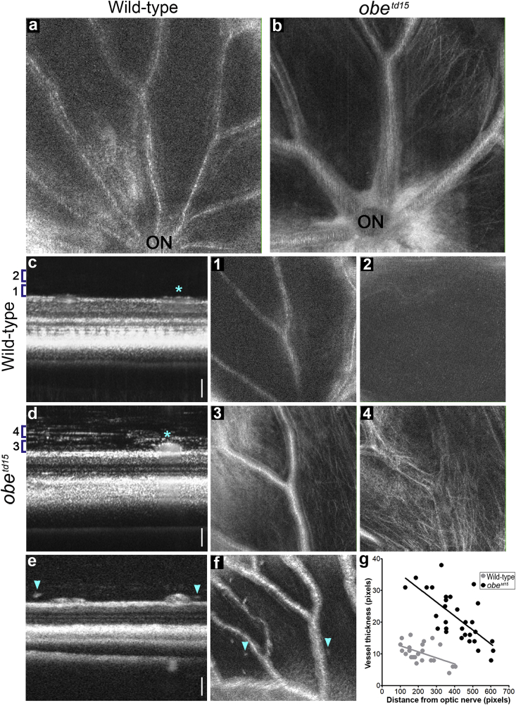
IMAGE

Fig. 4

ID
ZDB-IMAGE-200124-55
Source
Figures for Toms et al., 2019
Image
Figure Caption

Fig. 4

Retinal vasculature in obetd15 zebrafish.

OCT en face images of the dorsal retina show inner retinal vessels emerging from the optic nerve region in the wild-type (a) and obetd15 (b) fish at 12 months post-fertilization, displaying abnormal blood vessel appearance in the mutant retina. Cross-sectional B-scans from wild-type (c) and obetd15 (d) fish also demonstrate notable differences in vessel size (vessels indicated with *). Numbered brackets (1–4) on the cross-sections represent areas analyzed in the corresponding en face images. Fibrous material was apparent at each depth examined in the vitreous of the obetd15 retina (3, 4) but was not seen on the wild-type images (1, 2). Hyper-reflective deposits were also noted in the vitreous of the obetd15 retina on both B-scan (e) and en face (f) images, indicated with arrows. Graph (g) shows retinal vessel thickness plotted against distance from the optic nerve in wild-type and obetd15 zebrafish. Scale bars = 50 μm.

Figure Data
Acknowledgments
This image is the copyrighted work of the attributed author or publisher, and ZFIN has permission only to display this image to its users. Additional permissions should be obtained from the applicable author or publisher of the image.

Reprinted from Experimental Eye Research, 189, Toms, M., Dubis, A.M., Lim, W.S., Webster, A.R., Gorin, M.B., Moosajee, M., Missense variants in the conserved transmembrane M2 protein domain of KCNJ13 associated with retinovascular changes in humans and zebrafish, 107852, Copyright (2019) with permission from Elsevier. Full text @ Exp. Eye. Res.