IMAGE

Fig. 1 S1

ID
ZDB-IMAGE-191230-606
Source
Figures for Rahman et al., 2019
Image
Figure Caption

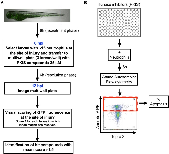
Fig. 1 S1 Schematics showing PKIS screen design.

(A) Tail fin transected three dpf Tg(mpx:GFP)i114 zebrafish larvae that had generated an inflammatory response at six hpi were incubated with individual PKIS compounds [25 µM] for a further 6 hr. Larvae were imaged and manually scored between 0–3 on the basis of green fluorescence at the injury site. (B) PKIS compounds were incubated with primary human neutrophils for 6 hr. Apoptosis was assessed by Annexin V/TO-PRO-3 staining by flow cytometry and the percentage apoptosis calculated as Annexin V single plus Annexin V/TO-PRO-3 dual positive events (as indicated by red box).

Acknowledgments
This image is the copyrighted work of the attributed author or publisher, and ZFIN has permission only to display this image to its users. Additional permissions should be obtained from the applicable author or publisher of the image. Full text @ Elife