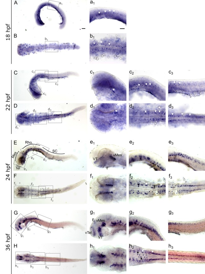
IMAGE

Figure 3

ID
ZDB-IMAGE-191230-54
Genes
Source
Figures for Lechermeier et al., 2019
Image
Figure Caption

Figure 3

Whole mount RNA in situ hybridization of slc2a3a at early embryonic stages [18–36 hours post fertilization (hpf)]. Pictures in the left column depict alternating lateral (A,C,E,G) and dorsal (B,D,F,H) views. Anterior is to the left. Higher magnifications (a1–h3) corresponding to boxes in (A–H). Arrowheads indicate examples of labeled cells or cell populations. Note staining in spinal cord, hindbrain, ventral midbrain, ventral thalamus, ventral telencephalon and epiphysis. Two bilateral rows, one medially (black arrowheads) and one laterally (white arrowheads) of positive cells are situated in the ventral hindbrain along the floor plate (f2 and h2). For abbreviations of anatomical terms see Table 1. Scale bars in (A), 100 μm and pertains to (A–H); scale bar in a1, 50 μm and pertains to a1–h3.

Figure Data
Acknowledgments
This image is the copyrighted work of the attributed author or publisher, and ZFIN has permission only to display this image to its users. Additional permissions should be obtained from the applicable author or publisher of the image. Full text @ Front. Mol. Neurosci.