IMAGE

FIGURE 1

ID
ZDB-IMAGE-191230-1140
Antibodies
Source
Figures for Vecchia et al., 2019
Image
Figure Caption

FIGURE 1

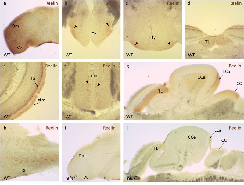
Reelin immunohistochemistry. Anti-Reelin antibody labeling of coronal and sagittal sections of the adult zebrafish brain. In wild-type anti-Reelin antibody labeling is seen in the (a) medial part of the dorsal telencephalon (Dm) and in the ventral nucleus of the ventral telencephalon (Vm), in the (b) ventral thalamic nuclei (Th) and (c) the ventral hypothalamic nuclei (Hy). Labeling is also detected in the (d) torus longitudinalis (TL), (e) stratum fibrosum marginale (sfm) and the stratum opticum (so) of the optic tectum. (f) A few labeled cells are observed in the interpeduncular nucleus (nin). In the hindbrain, Reelin protein localizes to the (g) corpus cerebelli (CCe), the lobus caudalis cerebelli (LCa), crista cerebellaris (CC) and (h) the intermediate and inferior part of the reticular formation (RF). (i,j) Anti-Reelin antibody labeling is not detected in the brain of reln–/– mutants. Black arrowheads show position of Reelin-positive areas in (b,c,f).

Figure Data
Acknowledgments
This image is the copyrighted work of the attributed author or publisher, and ZFIN has permission only to display this image to its users. Additional permissions should be obtained from the applicable author or publisher of the image. Full text @ Front. Behav. Neurosci.