
|
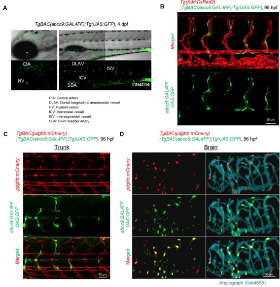
Fig. S1
TgBAC(abcc9:GAL4FF) zebrafish line. (A) Lateral view of the TgBAC(abcc9:GAL4FF);Tg(UAS:GFP) larva at 4 dpf. Upper, the merged image of bright field image and UAS:GFP; lower, UAS:GFP. (B-D) Lateral view of confocal stack image of trunk vasculature of TgBAC(abcc9:GAL4FF);Tg(UAS:GFP);Tg(kdrl:DsRed2) larva (B), trunk (C) or brain vasculature (D) of TgBAC(abcc9:GAL4FF);Tg(UAS:GFP);Tg(pdgfrb:mCherry) larva at 96 hpf. Lateral view, anterior to the left. Note that abcc9 reporter positive cells were hardly detected beneath the DA (B, C). In D, vascular structure was visualized by injecting Qdot655 into the circulation (aqua).