
Image
|
Figure Caption
Fig. S1
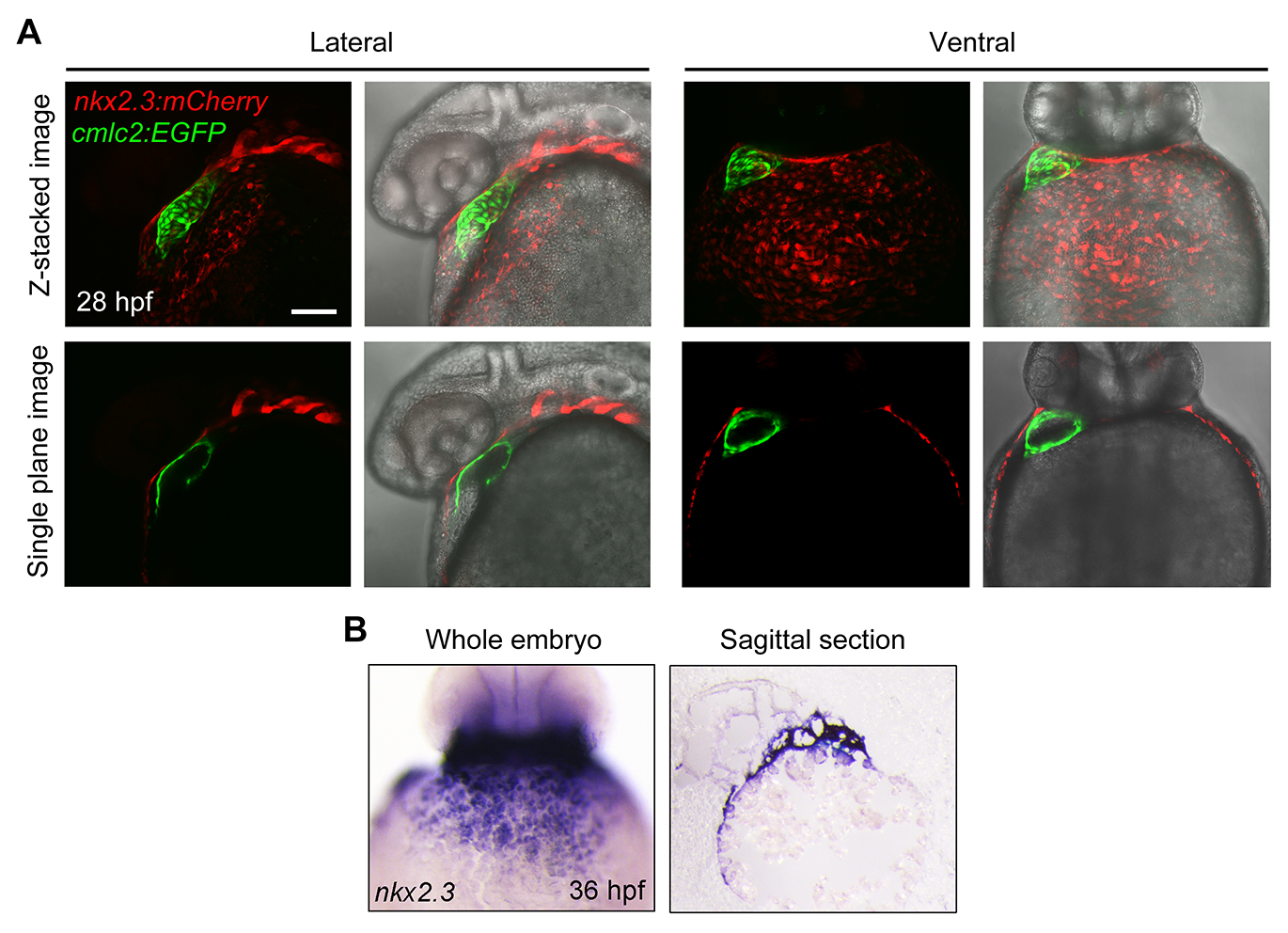
( A) Tg(nkx2. 3: mCherry;cmlc2: EGFP) embryos at 28 hpf with mCherry fluorescence (red) in the pericardium and EGFP fluorescence (green) in the heart. Single plane images showed that the GFP-positive myocardial cells were surrounded by the mCherry-positive cells. Scale bar, 50 μm. ( B) In situ hybridization of nkx2. 3 in whole mount embryos at 36 hpf (left panel, ventral view with anterior to the top). Sagittal section of the same embryo was shown in the right panel.
Acknowledgments
This image is the copyrighted work of the attributed author or publisher, and
ZFIN has permission only to display this image to its users.
Additional permissions should be obtained from the applicable author or publisher of the image.
Full text @ PLoS Genet.