
Image
|
Figure Caption
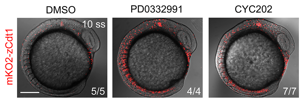
Fig. S8
Tg(mKO2-zCdt1(1/190)) embryos were treated with 0.5 μM PD0332991 or 0.2 μM CYC202 from the shield stage to the 10-somite stage. Then, these embryos were harvested for in vivo confocal imaging. Note that both PD0332991 and CYC202 treatments induced a remarkable increase of the number of mKO2-zCdt1–positive cells. Scale bar, 200 μm. CDK, cyclin-dependent kinase; EF1α, eukaryotic translation elongation factor 1α; mKO2, monomeric Kusabira Orange2; Tg, transgene; zCdt1, zebrafish chromatin licensing and DNA replication factor 1.
Acknowledgments
This image is the copyrighted work of the attributed author or publisher, and
ZFIN has permission only to display this image to its users.
Additional permissions should be obtained from the applicable author or publisher of the image.
Full text @ PLoS Biol.