
Image
|
Figure Caption
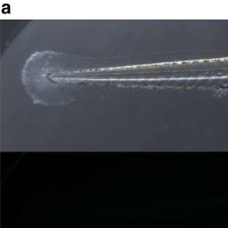
Fig. 1
Biodistribution after PSNP exposure in zebrafish larvae from 72 to 120 hpf. a Representative pictures of PSNP accumulation in the intestine (lateral view, solid arrow), exocrine pancreas (lateral view, empty arrow), and b gallbladder (ventral and lateral view, asterisk) of wild-type zebrafish larvae. The experiment was conducted on three separate occasions with ten biologically independent replicates each
Acknowledgments
This image is the copyrighted work of the attributed author or publisher, and
ZFIN has permission only to display this image to its users.
Additional permissions should be obtained from the applicable author or publisher of the image.
Full text @ Commun Biol