
|
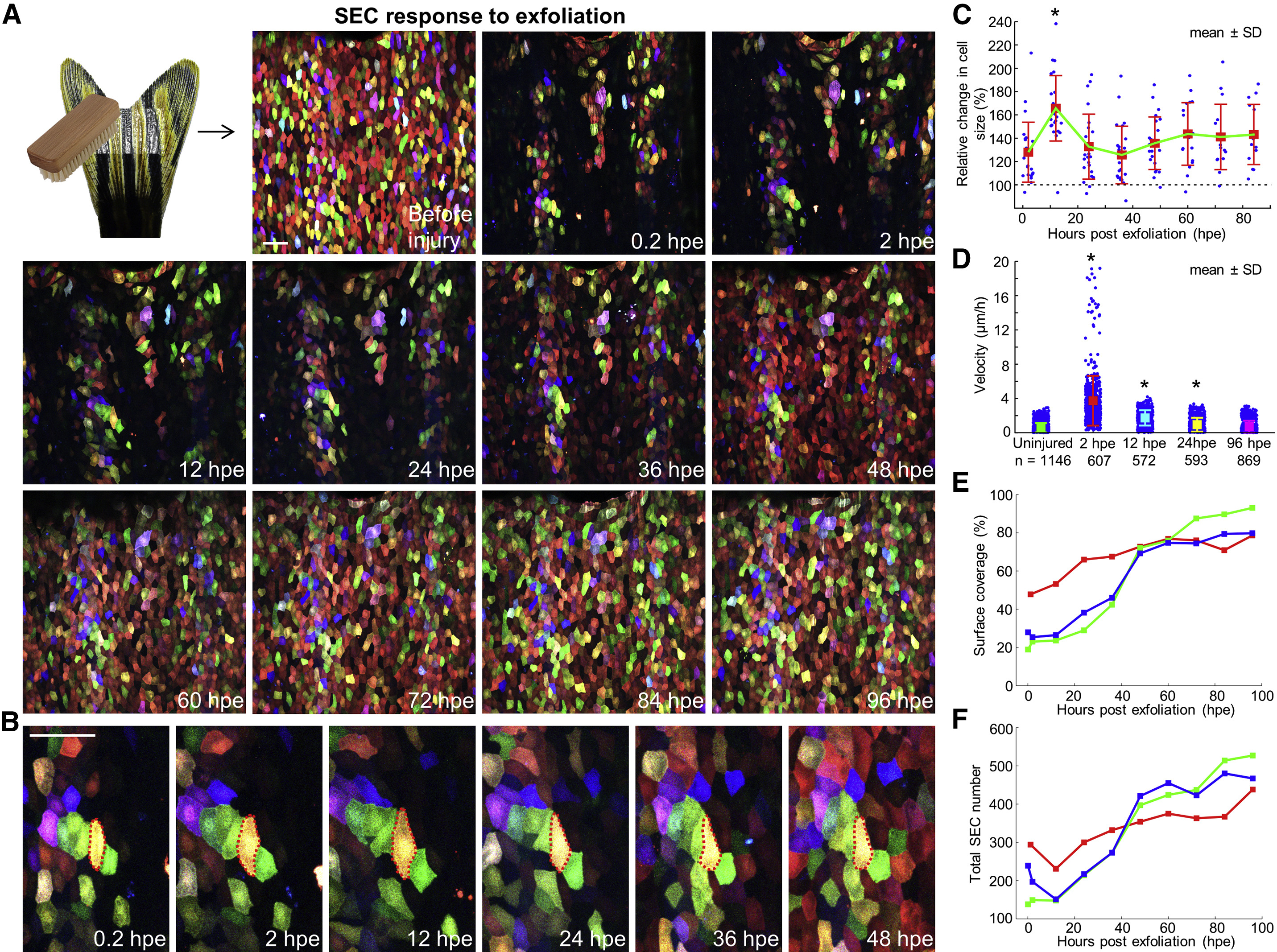
Fig. 4
Regeneration of Surface Injuries Involves a Biphasic Mechanism of Hypertrophy and Accelerated Replacement
(A) (Top left) Schematic of exfoliation injury. (Images) A time-lapse imaging series showing surface injuries and regeneration over a 4-day period. Scale bar, 50 μm.
(B) Enlarged view of a few cells during surface regeneration. Red dashed lines outline boundaries of a yellow cell undergoing transient hypertrophy. Scale bar, 50 μm.
(C) Quantification of relative change in SEC size after injury. The sizes of individual cells were normalized to their size immediately after injury, to indicate relative change (30% ± 5.4% greater at 2 hpe; 65.7% ± 5.9% greater at 12 hpe). Individual trajectories of spared SECs within the injured region were extracted from three independent scratch injuries (n = 20, 24, and 11). Bars indicate mean ± SD. Statistical significance was assessed by a Mann-Whitney test (2 hpe versus 12 hpe, ∗p < 10−4).
(D) Quantification of SEC velocities after injury. PIV type techniques were used to track single points in the images and compare typical velocities across adjacent time frames. Uninjured SECs and those at 96 hr post scratch (hps) show similar velocities, while SECs are more motile immediately after injury. Values below the plot are the number of points (blue) used to build the plot. Bars indicate mean ± SD. Statistical significance was assessed by a Mann-Whitney test (uninjured versus 2 hpe, 12 hpe, or 24 hpe, ∗p < 10−5 for all cases).
(E) Quantification of change in surface coverage during regeneration (n = 3).
(F) Quantification of change in total SEC number during regeneration (n = 3).
Reprinted from Developmental Cell, 36, Chen, C.H., Puliafito, A., Cox, B.D., Primo, L., Fang, Y., Di Talia, S., Poss, K.D., Multicolor Cell Barcoding Technology for Long-Term Surveillance of Epithelial Regeneration in Zebrafish, 668-80, Copyright (2016) with permission from Elsevier. Full text @ Dev. Cell