
Image
|
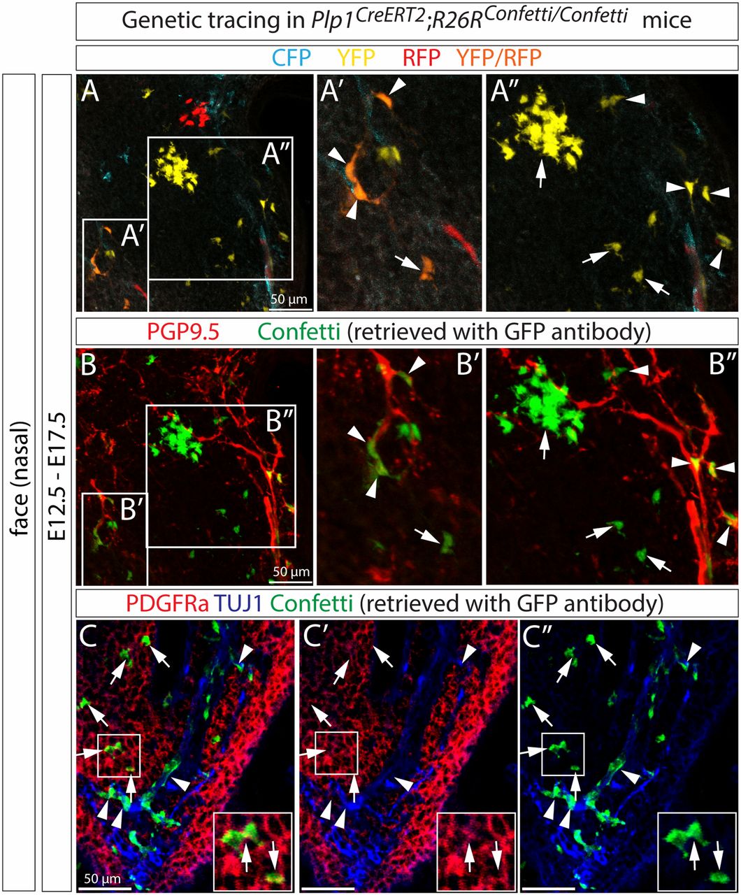
Figure Caption
Fig. 1
SCPs generate mesenchymal cells during murine embryonic development. (A–C) Genetic tracing of Plp1CreERT2;R26RConfetti/Confetti embryos revealed SCP contribution to proximal mesenchymal cells. Confetti clones expressing RFP, YFP, and/or CFP proteins are shown in A. The same tissue section was immunostained with PGP9.5 (for neuron) and GFP (for confetti) in B. (C) The traced cells off TUJ1+ nerves were positive for mesenchymal marker, PDGFRa. In A–C, arrowheads indicate traced cells on nerves and arrows indicate traced cells that become mesenchymal cells.
Acknowledgments
This image is the copyrighted work of the attributed author or publisher, and
ZFIN has permission only to display this image to its users.
Additional permissions should be obtained from the applicable author or publisher of the image.
Full text @ Proc. Natl. Acad. Sci. USA