
|
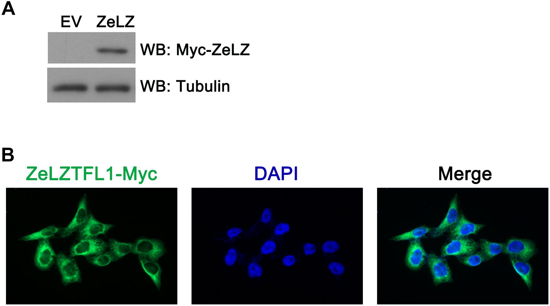
Fig. 5
(A) Myc-tagged zebrafish LZTFL1 plasmids were transfected into HeLa cells. At 48 h post-transfection, cells were lysed for Western blot analysis. As shown in the picture, compared with empty vectors, LZTFL1 fused with Myc tag was ectopically expressed in HeLa cells. (B) Myc-tagged zebrafish LZTFL1 was transfected into HeLa cells. At 48 h post-transfection, cells were fixed and probed with FITC-anti-Myc antibodies and DAPI (nuclear counterstain). Moreover, zebrafish LZTFL1 showed predominant localization in the cytoplasm but subtle localization in nuclei. Magnification, 200×. EV, empty vector, pcDNA6-Myc; ZeLZ, pcDNA6-Myc-ZeLZTFL1, zebrafish LZTFL1 was inserted into pcDNA6-Myc.