
|
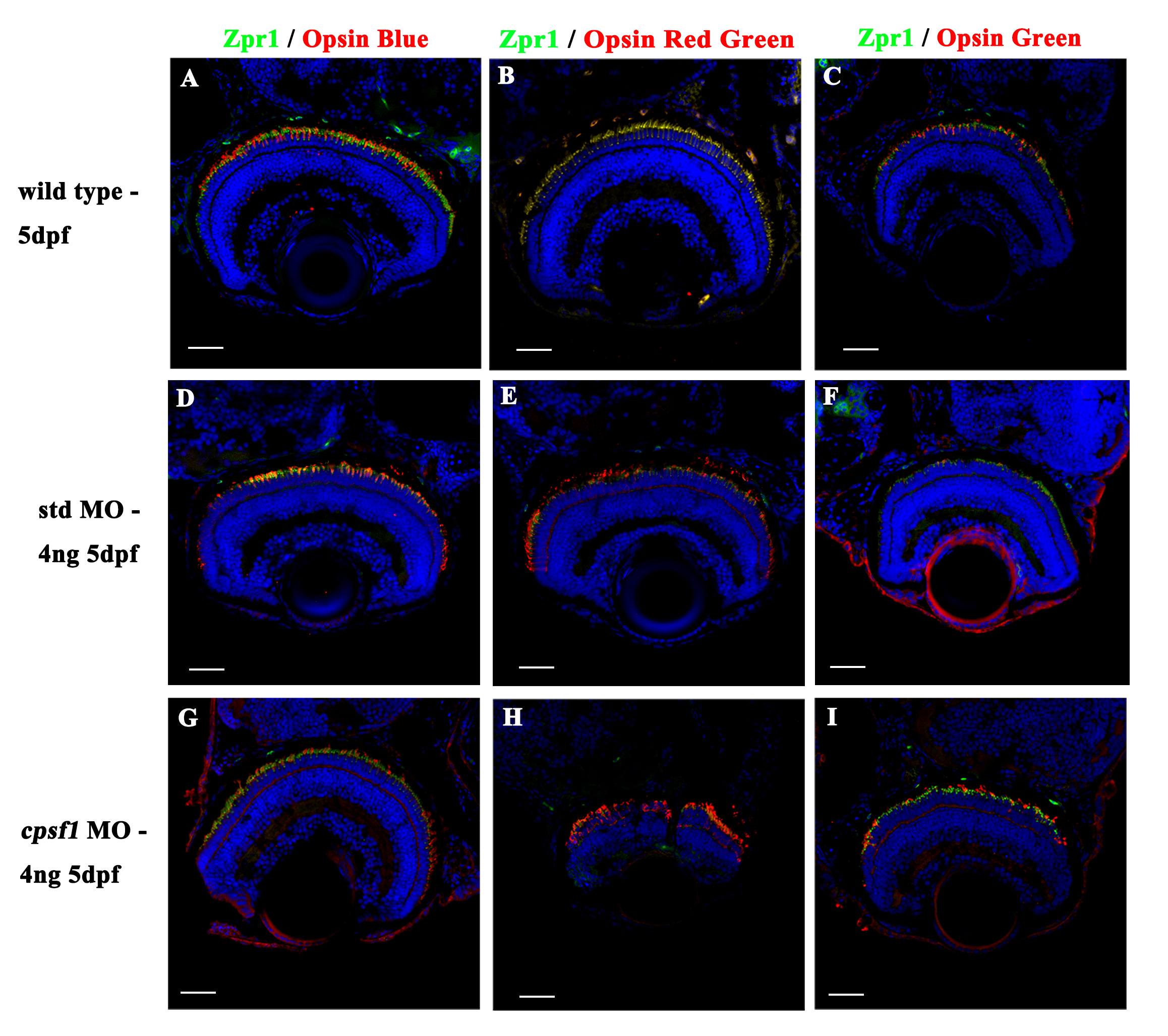
Figure S5
Photoreceptor cells developed normally in wild-type larvae (A-C), std MO larvae (D-F), and cpsf1 morphants (G-I). (A, D, G): Co-labelling with anti-Zpr1 (for double-cone photoreceptor cells, green) and anti-Opsin Blue (for blue cone photoreceptor cells, red) antibodies showed that the blue cone photoreceptor cells were mature. (B, E, H): Co-labelling with anti-Zpr1 (for double-cone photoreceptor cells, green) and anti-Opsin Red/Green (for red/green cone photoreceptor cells, red) antibodies indicated that the red and green cone photoreceptor cells developed normally. (C, F, I): Co-labelling with anti-Zpr1 (for double-cone photoreceptor cells, green) and anti-Opsin Green (for green cone photoreceptor cells, red) antibodies demonstrated that the green cone photoreceptor cells attained maturity.