
|
Fig. 4
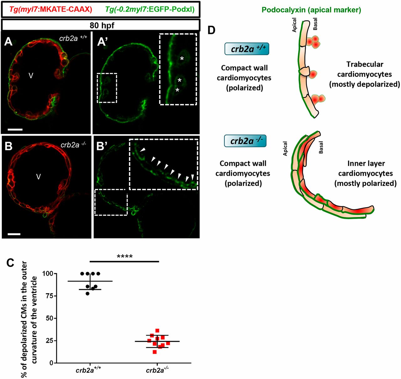
Most inner layer CMs in crb2a−/− remain polarized in the apicobasal axis. (A-B′) Confocal images (mid-sagittal sections) of 80 hpf Tg(−0.2myl7:EGFP-podxl); Tg(myl7:MKATE-CAAX); crb2a+/+ (A) and crb2a−/− (B) hearts. Asterisks in A′ mark delaminated CMs and arrowheads in B′ indicate polarized CMs. (C) Percentage of depolarized CMs in the outer curvature of the ventricle of 80 hpf crb2a+/+ (n=8) and crb2a−/− (n=10) larvae. Data are mean±s.e.m. ****P<0.0001 by Student's t-test. (D) Schematic illustration of A′ and B′ showing depolarized CMs in the trabecular layer of crb2a+/+ hearts and polarized CMs in the inner layers of crb2a−/− hearts. V, ventricle