
|
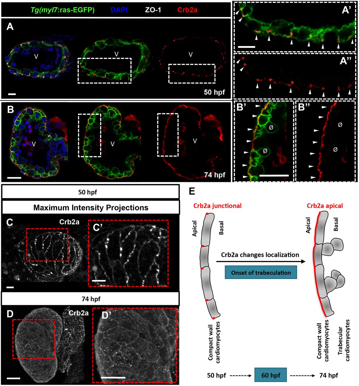
Fig. 1
Crb2a localization changes during cardiac trabeculation in zebrafish. (A-B″) Crb2a immunostaining in Tg(myl7:ras-EGFP)hearts at 50 (A-A″, n=12) and 74 (B-B″, n=15) hpf. At 50 hpf, Crb2a mostly localizes to the apical junctions between CMs (A-A″, arrowheads). At 74 hpf, Crb2a localizes to the entire apical membrane of compact layer CMs (B-B″, arrowheads), and is not observed in delaminated CMs (B′,B″, ø). (C-D′) Maximum intensity projections of ventricles showing junctional Crb2a immunostaining at 50 hpf (C,C′) and apical Crb2a immunostaining at 74 hpf (D,D′). (E) Schematic illustration of Crb2a relocalization in CMs from junctional to apical during the onset of trabeculation. V, ventricle.