IMAGE

Figure 1

ID
ZDB-IMAGE-190723-765
Source
Figures for Fontenas et al., 2018
Image
Figure Caption

Figure 1

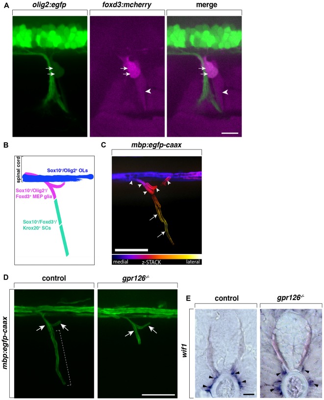
Centrally-derived motor exit point (MEP) glia myelinate motor nerve roots. (A) Lateral view of a olig2:egfp;foxd3:mcherry zebrafish trunk showing olig2+/foxd3+ MEP glia (arrows) and olig2/foxd3+ SCs (arrowheads) along a motor nerve at 55 hour post-fertilization (hpf). (B) Diagram representing central and peripheral myelinating glial markers. (C) Pseudo-colored z-stack of a lateral view of a mbp:egfp-caax trunk at 5 day post-fertilization (dpf). MEP glial myelin sheaths (arrowheads) originate within the spinal cord and project laterally along motor nerve axons, coming close to schwann cells (SC) myelin (arrows). (D) SC myelin (bracket) but not MEP glial myelin (arrows) is absent in G-protein coupled receptor 126 (gpr126) mutants at 5 days post fertilization (dpf). (E)In situ hybridization showing the presence of Wnt-inhibitory factor 1 (wif1+) MEP glia (arrowheads) along motor nerve roots in 3 dpf control and gpr126 mutant larvae. Scale bar, (A–D) 50 μm, (E) 20 μm.

Acknowledgments
This image is the copyrighted work of the attributed author or publisher, and ZFIN has permission only to display this image to its users. Additional permissions should be obtained from the applicable author or publisher of the image. Full text @ Front. Cell. Neurosci.