IMAGE

Fig 5

ID
ZDB-IMAGE-190723-319
Genes
Source
Figures for Blümel et al., 2019
Image
Figure Caption

Fig 5 Cryosections of the calvaria reveal precise localization of <italic>tcf12</italic>:EGFP-expressing cells.

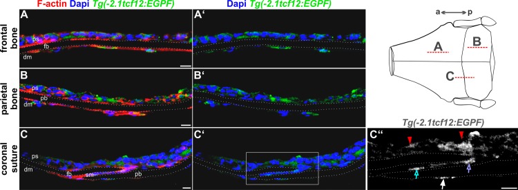
Confocal images of immunostaining on cryosections of the frontal and parietal bones and the coronal sutures of adult Tg(-2.1tcf12:EGFP) fish. Specimens were stained with anti-GFP (green) and counterstained with Phalloidin (F-actin, red) and DAPI (blue) for visualization of cell structures and nuclei. Sections of the frontal (A) and parietal (B) bones reveal tcf12:EGFP expressing cells in the periosteum and dura mater. (C) Section of the coronal suture unveils a GFP expression in the periosteum (red arrows in C”) and dura mater, too (white arrow in C”). A detail magnification (C”) unveils an additional GFP expression in the suture mesenchyme of the coronal suture (blue arrow in C”) and at the tip of the osteogenic front of the frontal plate (purple arrow in C”).The dashed lines in the scheme mark the area of the cryosections shown in A-C”. a, anterior; dm, dura mater; fb, frontal bone; p, posterior; ps, periosteum; pb, parietal bone; sm, suture mesenchyme. All scale bars represent 10 μm.

Figure Data
Acknowledgments
This image is the copyrighted work of the attributed author or publisher, and ZFIN has permission only to display this image to its users. Additional permissions should be obtained from the applicable author or publisher of the image. Full text @ PLoS One