
Image
|
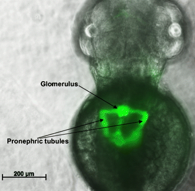
Figure Caption
Fig. 2
The developing kidney in a 48-h post-fertilisation zebrafish embryo. Shown is an image of a transgenic fish Tg(wt1b:EGFP), expressing the fluorescent protein E-GFP in kidney progenitor cells. The kidney is seen by its GFP expression, with notable structures labelled. The pronephric tubules have not yet reached their full length at this stage of development but will elongate further by 3.5 days post-fertilisation
Acknowledgments
This image is the copyrighted work of the attributed author or publisher, and
ZFIN has permission only to display this image to its users.
Additional permissions should be obtained from the applicable author or publisher of the image.
Full text @ Pediatr. Nephrol.