
|
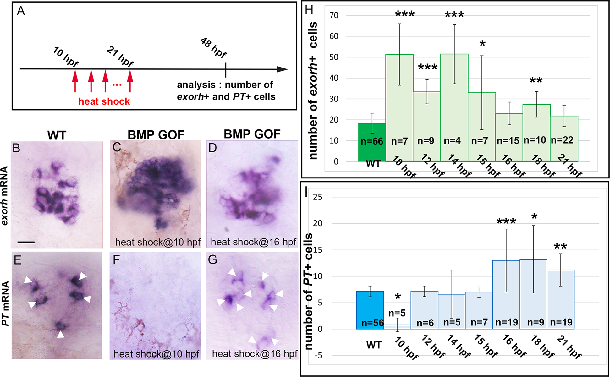
Fig. 7
(A) Scheme of the experimental design showing the timing of the heat shocks and analysis of the number of exorh+ and PT+ cells. (B-G) In situ hybridization for exorh and PT in 48-hpf WT and Tg(hsp70:bmp2b)fr13 transgenic embryos (BMP GOF) heat shocked at 10 or 16 hpf. Embryos are viewed dorsally with anterior to the top. Scale bar represents 10 μm. (H-I) Counts of exorh+ and PT+ cells in WT and Tg(hsp70:bmp2b)fr13embryos after a heat shock performed at various stages (indicated on the x-axis). Error bars represent SD. *p < 0.05. **p < 0.001. ***p < 0.0005. ****using a Kruskal-Wallis test with Dunn’s post hoc comparisons of the transgenic versus WT populations. Tg(hsp70:bmp2b)fr13 transgenic embryos were labeled BMP GOF. Underlying data can be found in S1 Data. BMP, bone morphogenetic protein; exorh, exorhodopsin; GOF, gain of function; hpf, hours post fertilization; PhR, photoreceptor; PT, parietopsin; WT, wild type.