
|
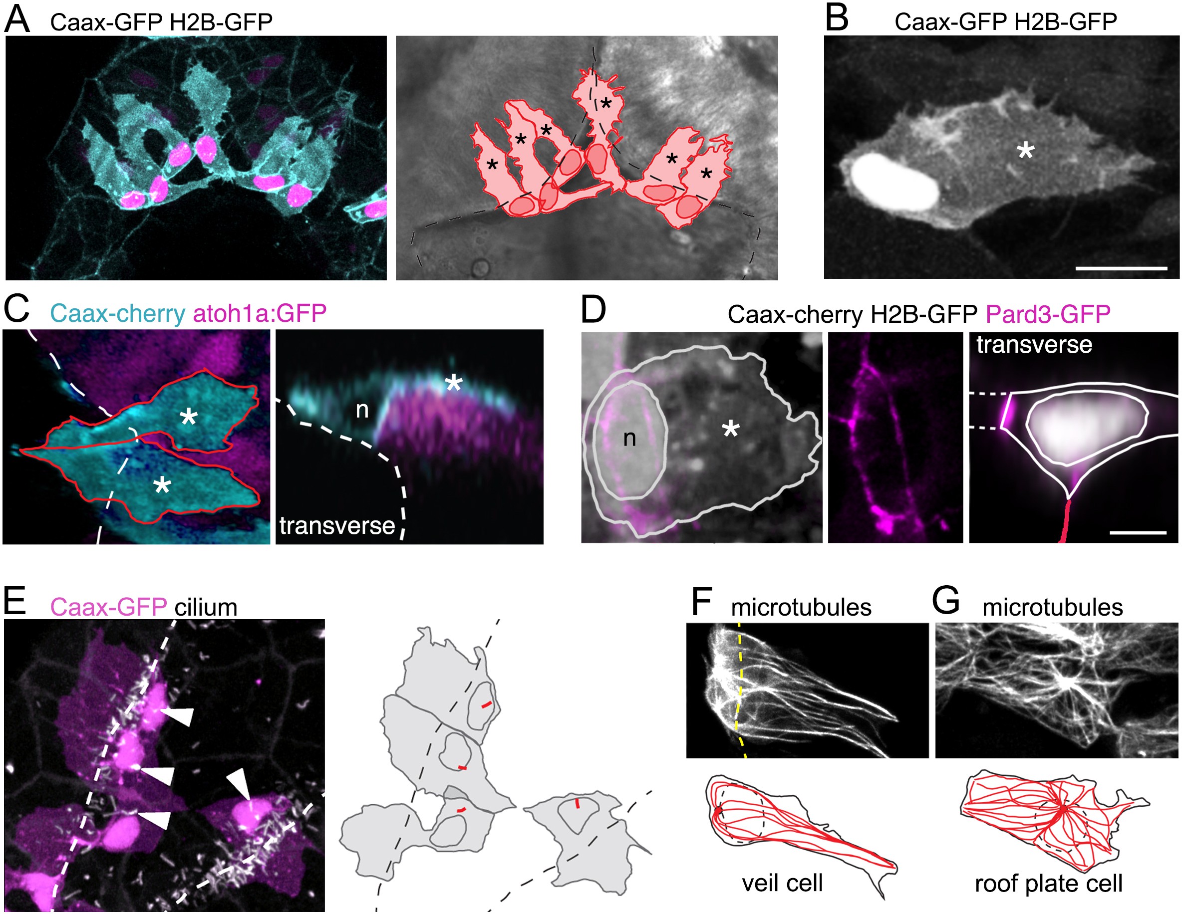
Fig. 3
A cell with novel morphology, the ‘veil’ cell, occupies the transition zone between squamous roof plate and columnar neuroepithelium.
(A) Dorsal view of six cells in the transition zone in the rostral pole of the hindbrain roof plate. Cells have distinctive morphologies with irregular lateral veils (asterisked in schematic to right) spanning the interface (dotted line) between roof plate and the columnar neuroepithelium. (B) Single veil cell with a nucleus placed at periphery of roof plate territory and a lateral veil (asterisk) extending over neuroepithelium. (C) Left. A dorsal view of two veil cells (cyan) with lateral veils (asterisks) extending over rhombic lip progenitors expressing atoh1a:GFP (magenta). Roof plate border shown with dashed line. Right. In transverse view reconstruction a veil cell presents a characteristically comma-shaped profile as its veil (asterisk) wraps itself over the basolateral surface of the atoh1-positive neural rhombic lip precursor. Apical surface of ventricle shown dashed, nucleus indicated with n. (D) Left: Dorsal view of veil cell, its nucleus (n) and veil (asterisk). Pard3-GFP expression shown in magenta. Middle: Single channel showing this cell’s apical ring of Pard3-GFP. Right: Same cell reconstructed in transverse plane showing Pard3-GFP (magenta) expression at the ventricular interface with squamous epithelium (dashed lines) and apical surface of columnar epithelium (red line). (E) Cilia (white) of veil cells (magenta) are located close to nuclei and protrude into ventricle. Roof plate border shown dashed. Many cilia from columnar cells are also visible in the maximum projection. (F) Two adjacent veil cells expressing Dck1k-GFP show highly polarized microtubule networks, comprising a near linear array extending into the veils. (G) By comparison, central cell in this image is a squamous roof plate cell displaying radially organized microtubule array around a centrally placed focal point.