
|
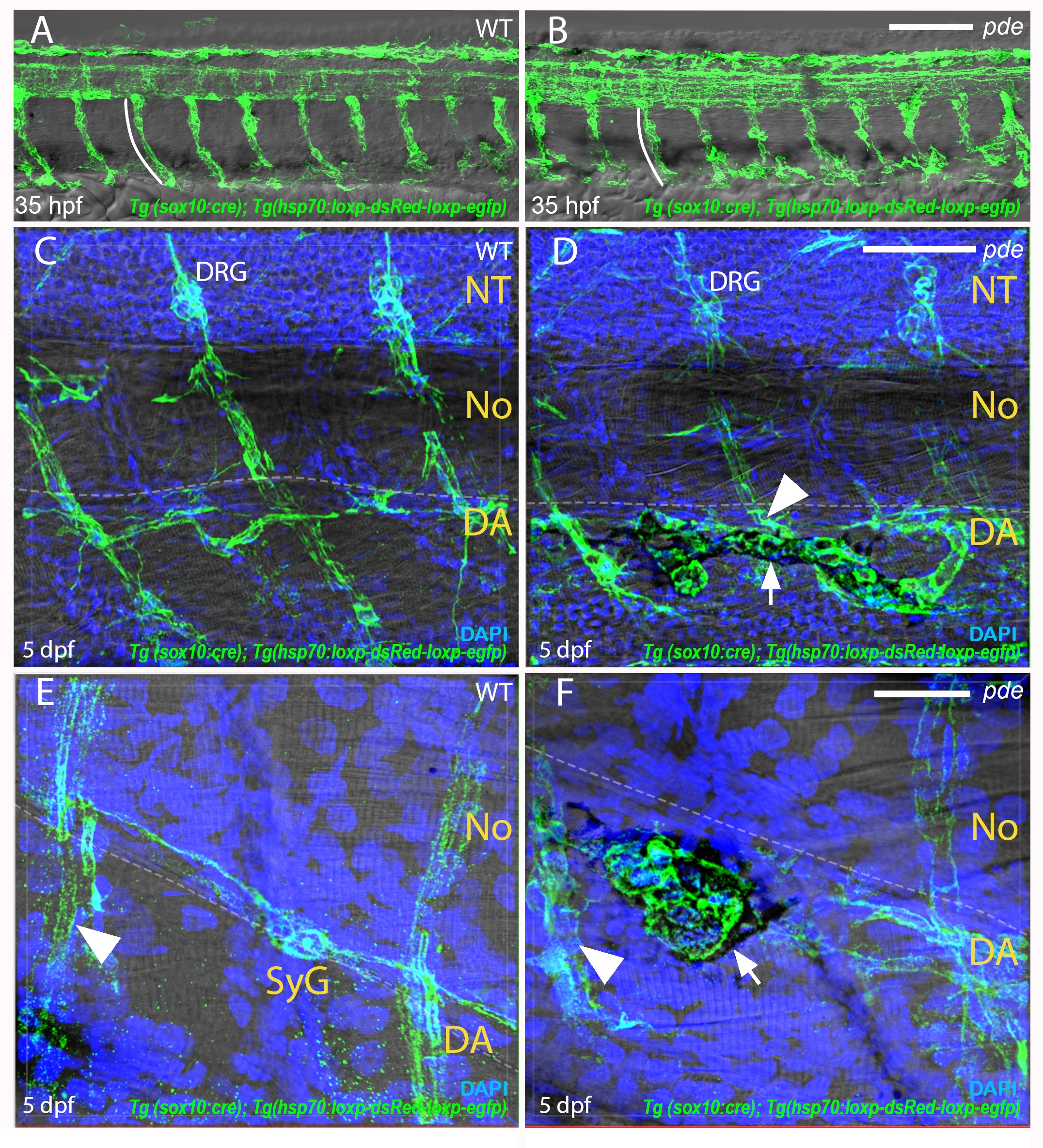
Fig. S3
Migration of neural crest cells through the medial migratory pathway.
Labelling of neural crest derivatives with GFP using the transgenic line Tg(-4725sox10:cre)ba74; Tg(hsp:loxp-dsRed-loxp-LYN-EGFP) shows no difference between 35 hpf WT fish (A) and pde mutants (B), neural crest cells migrate ventrally in a intersegmental arrangement (white line in A and B). 5 dpf pde mutant larvae show ectopic pigment cells (white arrow in D) associated with the spinal nerve projections (arrowheads in D) that emerge from the dorsal root ganglia (DRG). Ectopic pigment cells (white arrows) are also associated with the sympathetic ganglion (SyG) chain that forms perpendicular to the spinal nerve projections (white arrowhead in E and F) and ventral to the notochord (No). Guided by DIC image, dorsal edge of the dorsal aorta (DA) is highlighted with a dashed white line in C-F. Neural tube (NT). DAPI labels nuclei (blue). Scale bar = 25 μm (A and B), 50 μm (C and D) and 15 μm (E-F).