
Image
|
Figure Caption
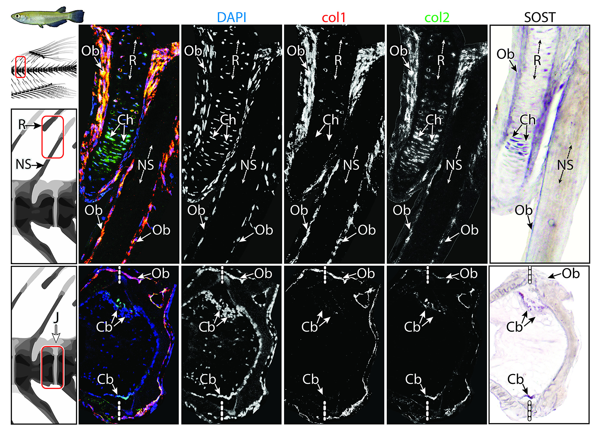
Fig. S4
Top row: NS and adjacent fin R; bottom row: intervertebral J. Each row comprises (from left to right) a multichannel RGB double fluorescent ISH image, followed by isolated single-channel images (DAPI, col1, col2, respectively) and a SOST ISH image. The figure provides a deconstruction of the multichannel ISH images Fig 4. Note SOST-positive Cbs, Chs, and Obs and the lack of osteocytes within the NS. Refer to text for further results. Cb, chordoblast; Ch, chondrocyte; ISH, in situ hybridization; J, joint; NS, neural spine; Ob, osteoblast; R, radial; RGB, red-green-blue.
Acknowledgments
This image is the copyrighted work of the attributed author or publisher, and
ZFIN has permission only to display this image to its users.
Additional permissions should be obtained from the applicable author or publisher of the image.
Full text @ PLoS Biol.