
|
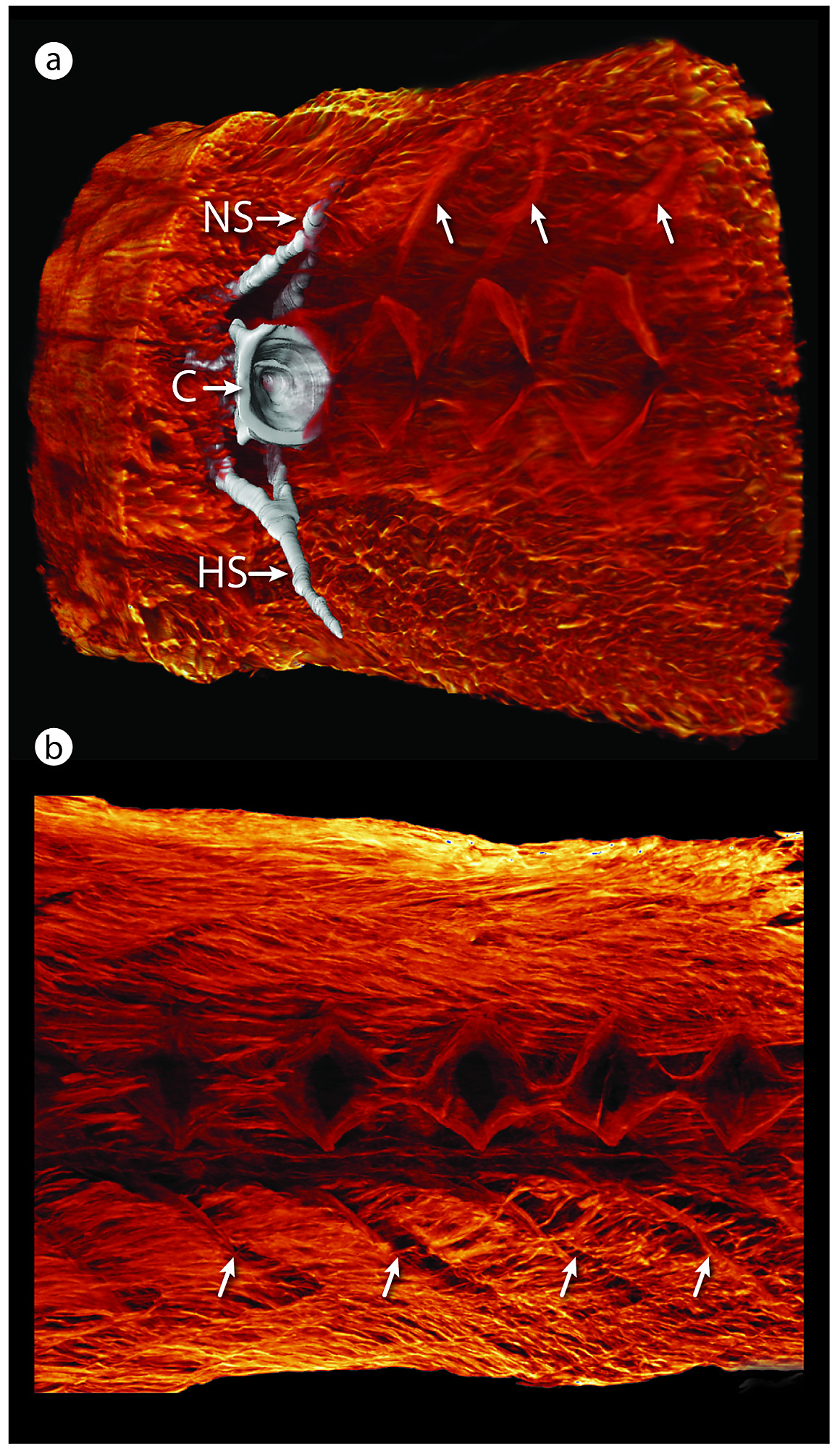
Fig. S1
Caudal is to the right in all images. (a) Sagittal section of a 3D reconstruction of contrast-enhanced (PTA) tomography showing the dense musculature surrounding the caudal vertebral column of medaka. One vertebra is segmented in white to show the full 3D muscle–vertebral relationship. The caudal faces of the vertebral C, NS, and HS are indicated. Neural arch muscular attachments are marked by white arrows. (b) Medial view of a sagittal section of medaka caudal musculature, with vertebrae digitally removed. The paravertebral musculature is complex and robust and occupies a large portion of the caudal part of the fish body, attaching mainly to the NS and HS and arches. Muscle fiber orientations and sites of attachment are clearly visible: intervertebral joints occupy the serial, diamond-shaped gaps in the musculature, and hemal arch muscular attachments are marked by white arrows. C, centrum; HS, hemal spine; NS, neural spine; PTA, phosphotungstic acid.