
|
Fig. 6
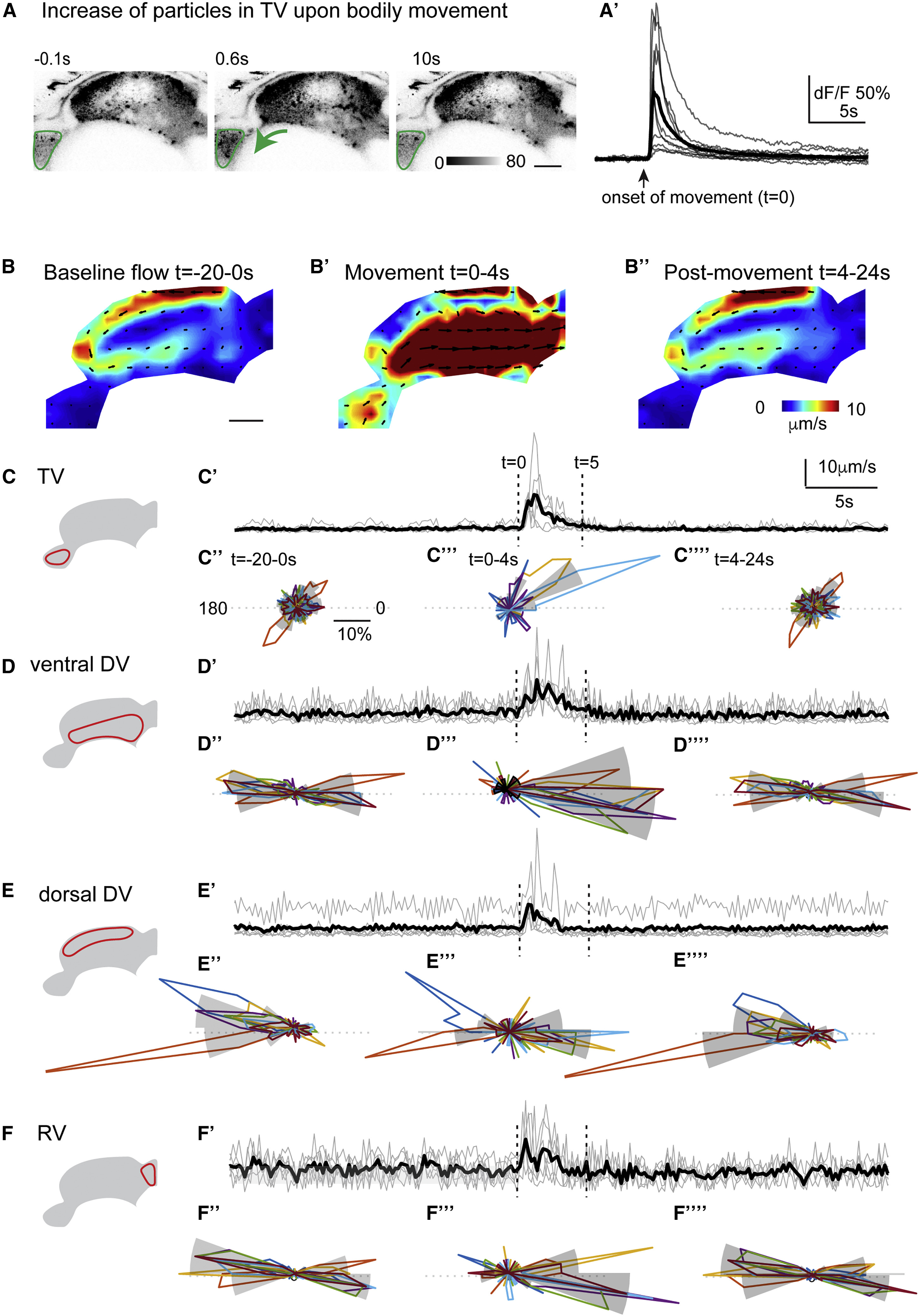
Upon Bodily Movement, the Stringent Compartmentalization of the Larval Ventricular System Is Temporarily Eliminated
(A–A’’) Upon bodily movement, the amount of fluorescent particles transiently increases in the telencephalic ventricle (TV), as shown by confocal microscopy of a fluorescent particle-injected larval brain before (−0.1 s), shortly after (0.6 s), or 10 s after bodily movement at 2 dpf (A). The onset of movement corresponds to time 0. The change of fluorescence (dF/F) in the TV (encircled in green in A) upon a single body contraction reveals a fast increase of fluorescence upon bodily motion followed by a slow return to baseline levels (A’). The average (n = 7 movements) is indicated in black.
(B–B’’) PIV analysis of fluorescent particle recordings indicates that the CSF flow fields are strongly influenced by movement at 2 dpf. Prior to bodily movement (t = −20–0 s), the directional CSF flow along the ventricular walls is evident, referred to as the baseline flow (B). After the bodily movement (t = 0–4.0 s), a strong surge of CSF backward from the TV to the rhombencephalic ventricle (RV) is prominent (B’). About 4.0 s (t = 4.0–24 s) later, the baseline flow as seen in (B) is re-established (B’’). Representative example of n = 10 is shown.
(C–F””) In order to quantify the impact of bodily movement on the CSF flow kinetics, we measured the flow velocity and the flow direction in all consecutive PIV-analyzed frames in the TV (C–C’’’’), the ventral DV (D–D’’’’), the dorsal DV (E–E’’’’), and the RV (F–F’’’’). The velocity of CSF flow increases transiently in the TV, DV, and RV following bodily movement, indicated by a dashed line at time 0, and returns to baseline after 5 s. Black is average of all the movement-induced flow (C’–F’).
(C’’–F’’’’) Bodily movements affect the directionality of CSF flow across the ventricles, as indicated by polar histograms of multiple frames preceding movement (−20–0 s; C’’–F’’), shortly after movement (0–4 s; C’’’–F’’’) and following movement (4–24 s; C’’’’–F’’’’). In the TV, the polar histograms show little flow directionality before movement (C’’), a strong directionality of 30° pointing caudally toward the DV shortly after movement (C’’’), and a re-establishment of baseline flow directionality 4 s after the bodily movements (C’’’’). In the ventral DV and RV, the CSF flow is highly pulsatile before and after bodily movement, as indicated by the axial distribution of flow direction pointing toward 180° and 0° (D’’ and F’’). Just after the movement, the flow is primarily oriented caudally toward 0° (D’’’ and F’’’). In the dorsal DV, the flow direction is oriented rostrally with an angle of 180° (C’’ and C’’’’), but after bodily movements, the flow is directed caudally (C’’’).
Scale bars are 50 μm. Each individual movement is represented (n = 7 movements). Mean is shown in gray. See also Figure S7 and Video S7.