
|
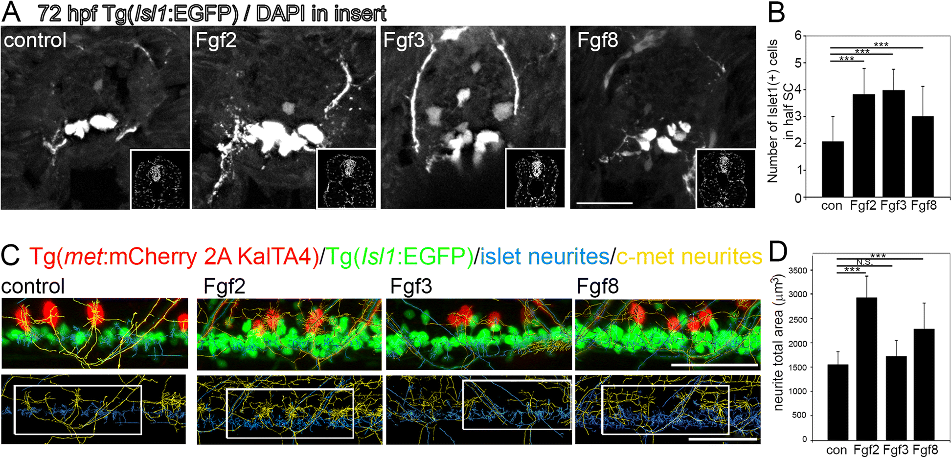
Fig. 6
Fgfs mediate neurogenesis and neurite outgrowth during zebrafish development at three days postfertilisation. a Tranverse sections through Tg(Isl1:GFP) spinal cords after 48 h incubation in Fgf2, 3 or 8, showing Islet1+ motor neurons. Insets show DAPI nuclear labelling in lower right corner for each image. b Quantitation of half spinal cord in the sections at the level of the back-fin shows a significant increase in Islet1+ motor neurons following incubation in Fgf2, 3 or 8. Results are presented in B as mean ± SEM, (n = 10 fish/group)*** p < 0.001. c Representative images of longitudinal spinal cord images of double transgenic Tg(Isl1:GFP)/ Tg(c-met:mCherry) fish incubated for 48 h in Fgf2, 3 or 8. Upper panel shows Islet1+ (green) and c-Met+ (red) transgenic label with Islet+ neuritis computationally annotated by Imaris software traced in blue and c-Met neurites computationally annotated by Imaris software traced in yellow. Lower panel shows an example of region of interest taken for analysis. d Quantitation of neurite total area of Islet1+ GFP and c-Met+ mCherry neurites reveals a significant increase in neurite outgrowth following Fgf2 and to a lesser extent Fgf8, but not Fgf3 incubation. Results are presented in D as mean ± SEM, (n = 8 fish/group) *** p < 0.001, N.S. = not significant. Scale bar in A is 50 μm, scale bar in C is 50 μm