
|
Fig. 8
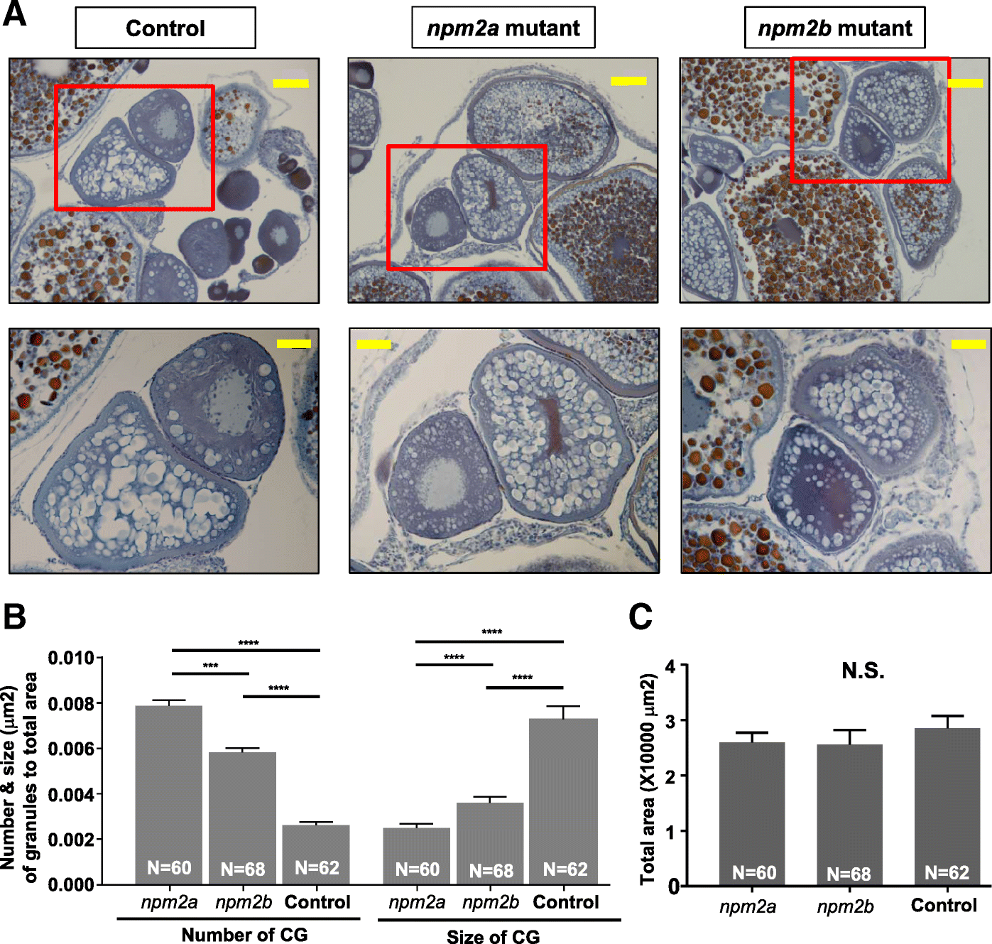
Histological analyses of the ovaries from nucleoplasmin (npm) 2a and npm2b mutant animals. a Ovary sections from age-matched npm2a, npm2b, and wildtype (WT) control females stained with Regaud’s hematoxylin. Top panels, 20X magnification; bars denote 90 μm; bottom panels show higher magnification images of the boxed areas in the top panels, 40X magnification; bars denote 45 μm. b Quantitation of the number and size (μm) of the cortical granules after adjusting to the total area of stage II follicles from ovaries from age-matched npm2a, npm2b, and control females. The N number denotes the total number of follicles counted for each group. c Total area of the stage II follicles from ovaries from age-matched npm2a, npm2b, and control females. N = 3 for all samples. N.S. = not significant, ***p < 0.001, ****p < 0.0001 by Mann-Whitney U-test