
Image
|
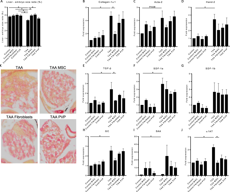
Figure Caption
Fig. 6
MSCs prevent the progression of liver fibrosis in zebrafish embryos. Quantitative PCR for mRNA expression of fibrotic, tissue damage and liver function genes after TAA treatment and MSC, Fibroblast or PVP injections. (A) At 8dpf the embryos were imaged to measure the sizes of the liver and total embryo in order to calculate the liver to embryo size ratio (N = 2, ±SEM). (B–J) Expression levels of Collagen1α1, Acta-2, Hand-2, TGF-β, SDF-1a, SDF1-b, GC, SAA and α1AT are normalized to RPP and to heathy control embryos. The graphs represent values of three independent experiments (n = 3, ±SEM). *p ≤ 0.05.
Acknowledgments
This image is the copyrighted work of the attributed author or publisher, and
ZFIN has permission only to display this image to its users.
Additional permissions should be obtained from the applicable author or publisher of the image.
Full text @ Sci. Rep.