
Image
|
Figure Caption
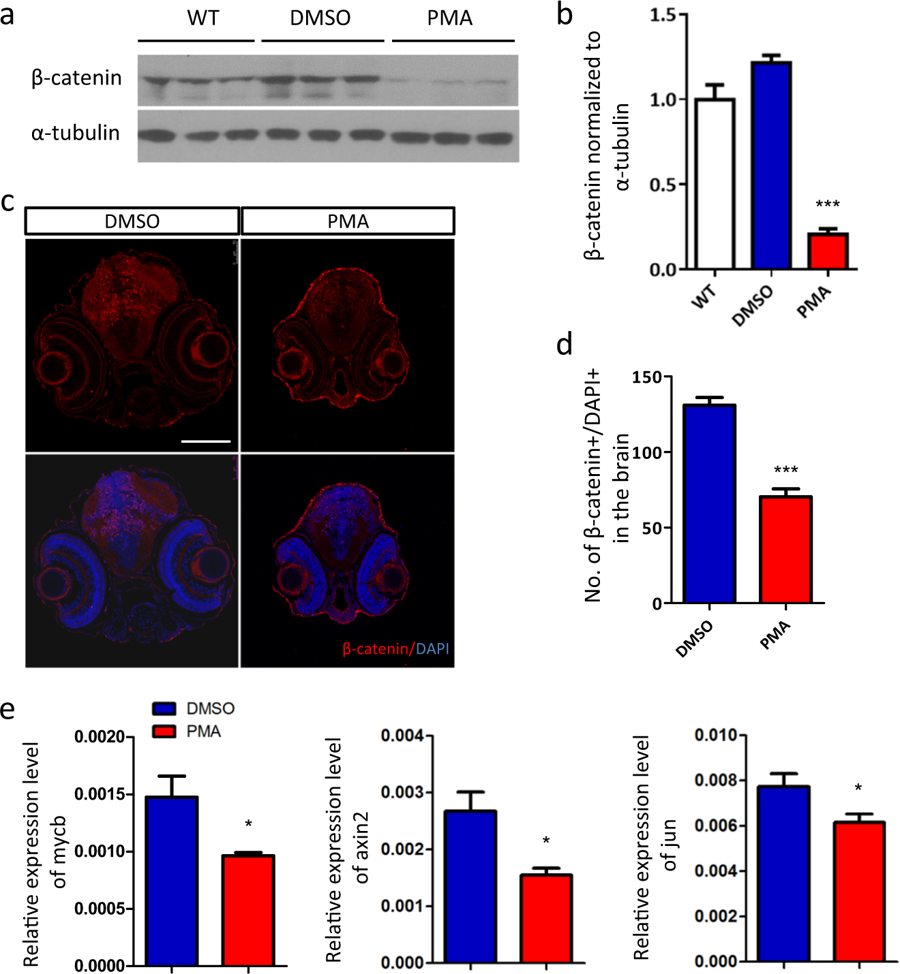
Fig. 3
Excessive PKC signaling promotes the degradation of β-catenin.
a Western blot analysis of β-catenin. b Quantification of the protein level of β-catenin relative to wild-type (WT) group (n = 3 per group, ***p < 0.001, t-test). c The nuclear accumulation of β-catenin is reduced by PMA treatment, d indicated by the quantification of β-catenin+/DAPI+ dots in brain region (n = 6 per group, ***p < 0.001, t-test). e RT-qPCR analysis of the transcriptional target of β-catenin, mycb, axin2 and jun (n = 6 per group, *p < 0.05, t-test)
Acknowledgments
This image is the copyrighted work of the attributed author or publisher, and
ZFIN has permission only to display this image to its users.
Additional permissions should be obtained from the applicable author or publisher of the image.
Full text @ Transl Psychiatry