
Image
|
Figure Caption
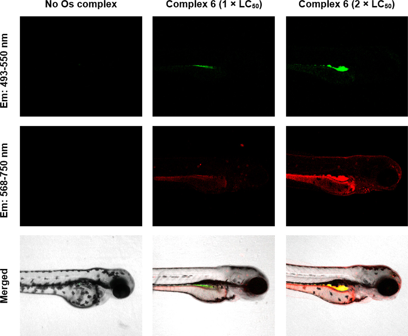
Fig. 6
Two-color fluorescent imaging of whole mount SG-WT zebrafish (Danio rerio) treated with Os(II) sulfonamide 6 for 96 h. Fluorescence for ROS (green) and 6 (red) is shown superimposed onto bright field images. Overlapping regions (yellow) are shown. Confocal images were acquired using a Zeiss LSM880 confocal microscope. Embryos were stained using the green reagent of the ROS/Superoxide Detection Kit (Enzo life sciences) for ROS detection. Excitation, 458, 488, and 561 nm; green emission for ROS, 493–550 nm; red emission for 6, 568–750 nm. See SI for full confocal data.
Acknowledgments
This image is the copyrighted work of the attributed author or publisher, and
ZFIN has permission only to display this image to its users.
Additional permissions should be obtained from the applicable author or publisher of the image.
Full text @ J. Med. Chem.