
|
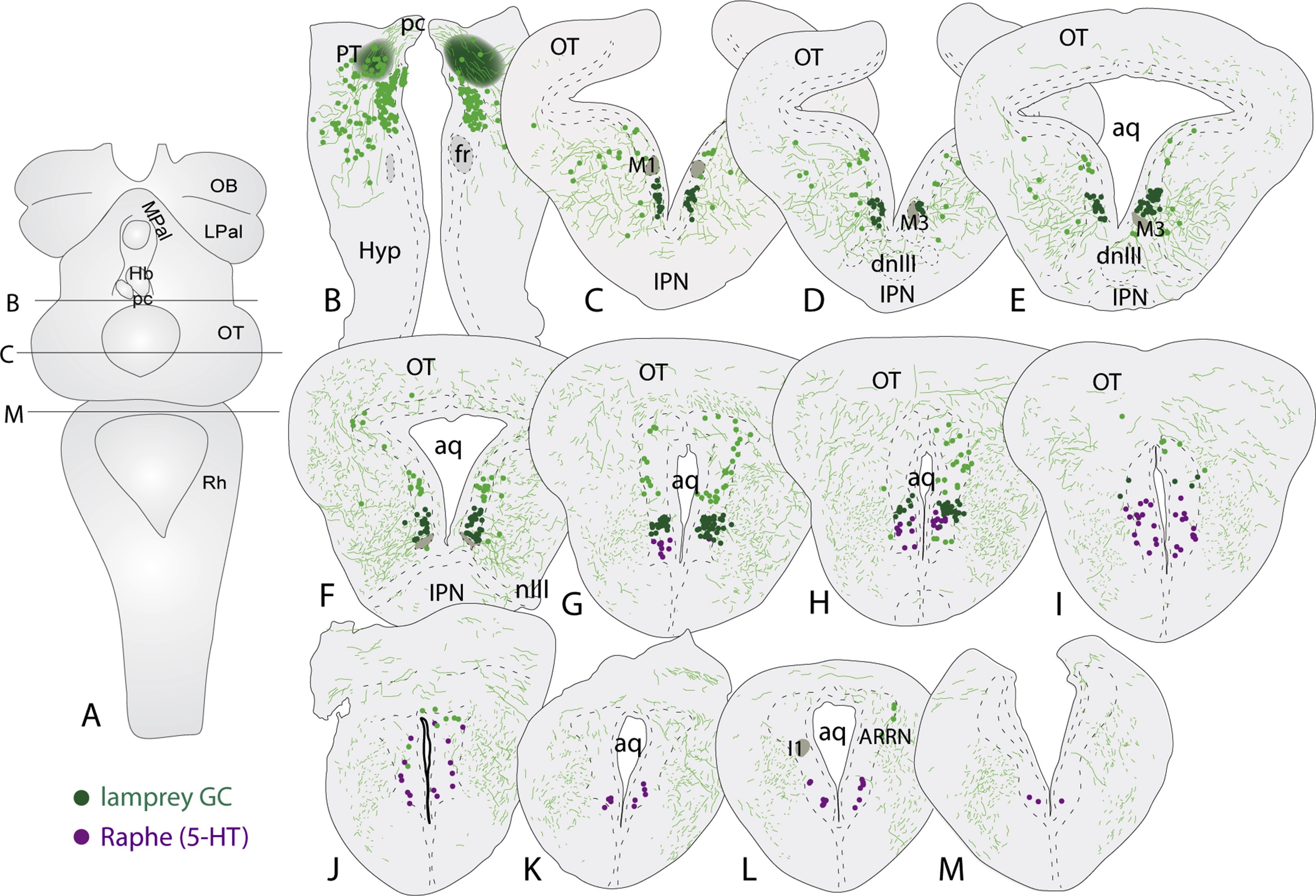
Fig. 3
Location of the lamprey griseum centrale. (A) Dorsal view of the lamprey brain indicating the level of the injection site (B), as well as the most rostral (C) and the most caudal section (M). (B) The bilateral injection sites in the pretectum. (C–M) Schematic drawing illustrating the rostrocaudal extent of griseum centrale (GC; green) and its relation to the serotonergic raphe nucleus (magenta). Aq, aqueduct; ARRN, anterior rhombencephalic reticular nucleus; dnIII, dorsal nucleus of the oculomotor nerve; Hyp, hypothalamus; I1, I1 Müller cell; IPN, interpeduncular nucleus; nIII, the oculomotor nerve; OT, optic tectum; pc, posterior commissure; PT, pretectum. (For interpretation of the references to colour in this figure legend, the reader is referred to the web version of this article.)