
|
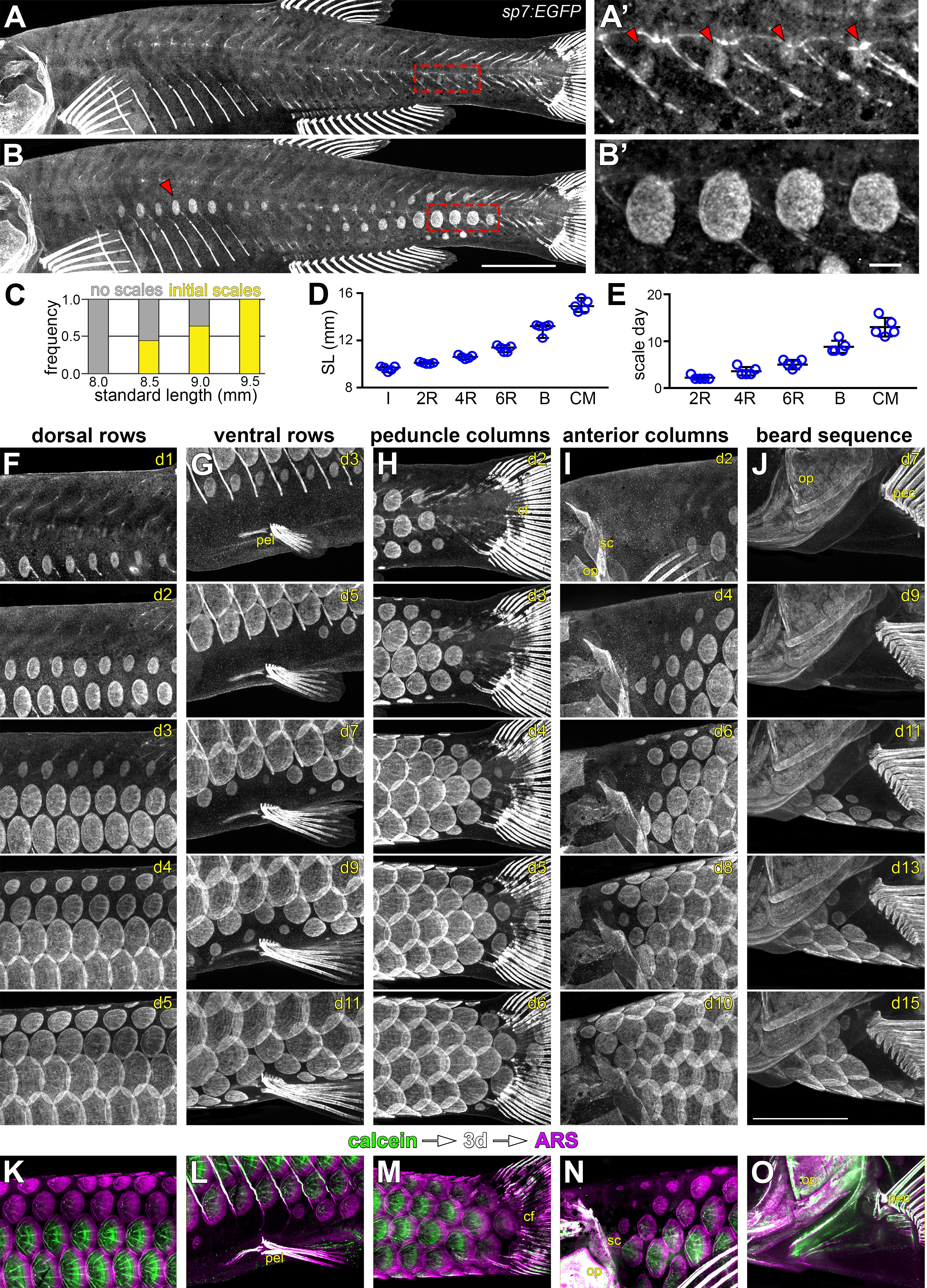
Fig. 1-S2
Squamation details revealed by repeated live imaging of individual fish.
(A,B) Squamation initiated from two foci, on the caudal peduncle (boxed in a), and on the flank above the ribs (red arrowhead in B). (A’,B’) Closer view of boxed areas in (a) and (b). (C) Squamation initiated in larvae between 8.5 mm and 9.5 mm standard length (SL; n = 131). (D,E) Squamation was completed over 13 ± 2 days during a period of rapid somatic growth. Measurements shown are from five representative individuals housed individually, fed ad libitum and imaged daily throughout squamation. (F,G) Addition of scale rows and columns in a single representative individual. (K–O) Sequential calcein, ARS labelling confirms squamation sequence revealed by daily live imaging. pel = pelvic fin; cf = caudal fin; op = opercle; so = superopercule bone; pec = pectoral fin. Scale bars, 1 mm (A, B, F–O); 100 µm (A’, B’).