
Image
|
Figure Caption
Fig. S3
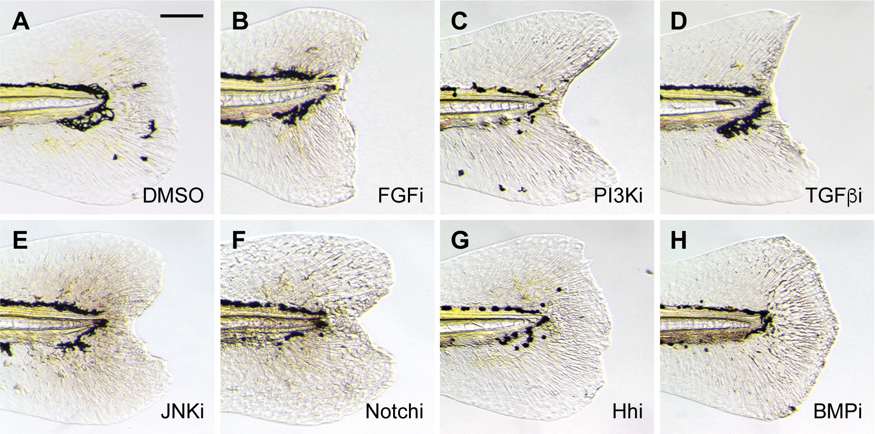
Inhibition of larval tail regeneration by pathway-specific antagonists.
Representative micrographs of larval tails that were amputated at 2 dpf and then treated with the following signaling pathway inhibitors for 3 days: (A) 0.5% DMSO; (B) 75 μM PD173074 (FGF); (C) 10 μM LY294002 (PI3K); (D) 50 μM SB431542 (TGFß); (E) 5 μM SP600125 (JNK); (F) 50 μM DAPT (Notch); (G) 100 μM cyclopamine (Hh); or (H) 50 μM dorsomorphin (BMP). At least 30 larvae were analyzed for each experimental condition, and phenotypic descriptions were based on a penetrance of > 80%. Scale bar: 100 μm.
Acknowledgments
This image is the copyrighted work of the attributed author or publisher, and
ZFIN has permission only to display this image to its users.
Additional permissions should be obtained from the applicable author or publisher of the image.
Full text @ PLoS One