
|
Fig. 1
Formation of Shh-Dependent Muscle Requires RyR Function
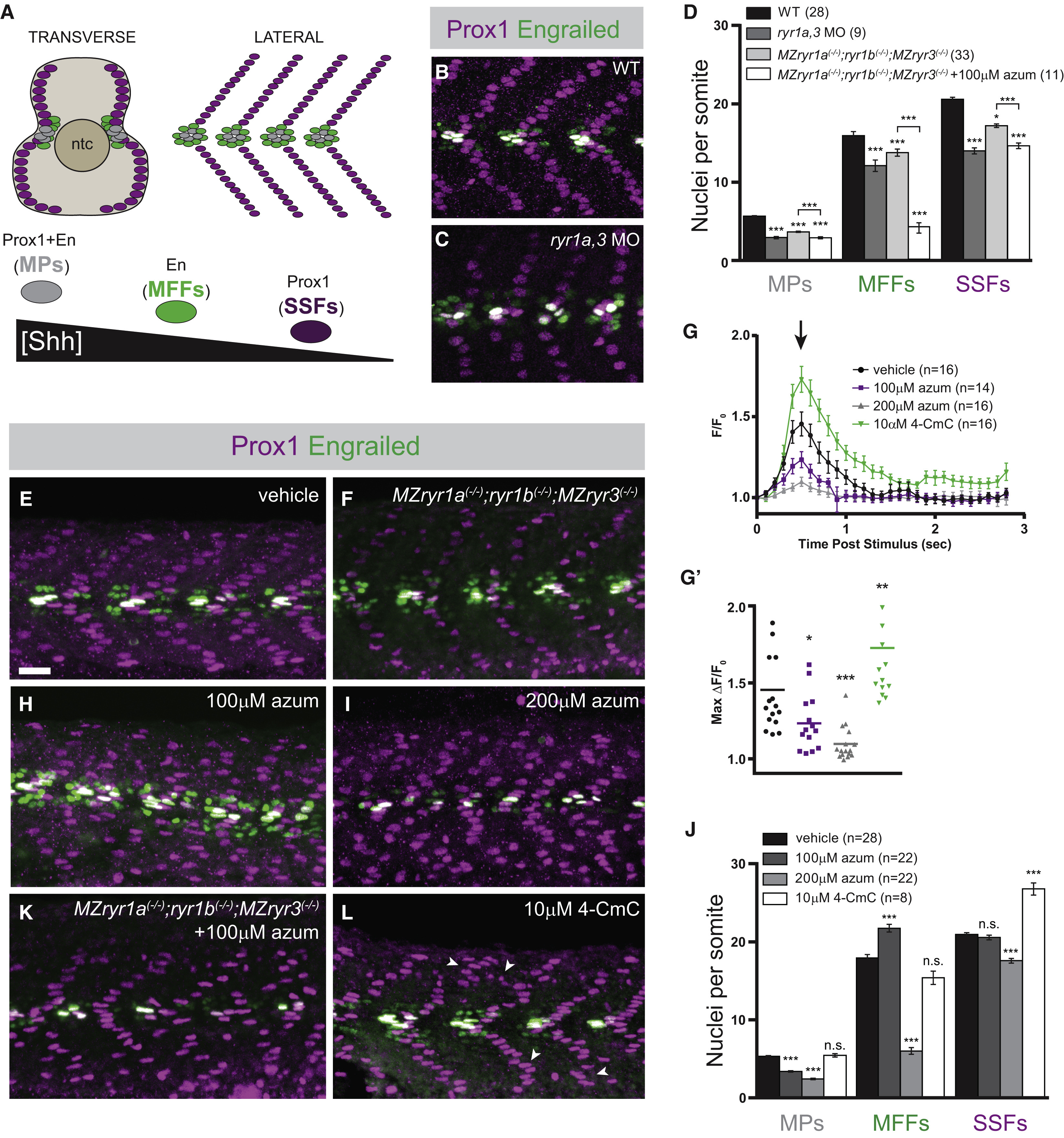
(A) Schematic illustration of the nuclei of Shh-dependent muscle cells in the somites of 26 hpf zebrafish embryos, presented relative to the notochord (ntc) in transverse section and in a superficial lateral view (anterior to left). Nuclei of different muscle types are color-coded: slow muscle pioneer cells ([MPs], gray), medial fast fibers ([MFFs], green), and superficial slow fibers ([SSFs], magenta).
(B, C, E, F, H, I, K, and L) Shh-dependent muscle fiber nuclei, marked by Prox1 (magenta) and En (green) expression, were visualized by immunostaining 26 hpf embryos: (B) wild-type (WT), (C) ryr1a and ryr3 MO-injected (ryr1a,3 MO), (E) vehicle-treated (0.5% DMSO) WT, (F) MZryr1a(−/−);ryr1b(−/−);MZryr3(−/−) mutant, (H and I) azumolene-treated WT, (K) azumolene-treated MZryr1a(−/−);ryr1b(−/−);MZryr3(−/−) mutant, and (L) 4-CmC-treated WT (supernumerary Prox1+ SSF nuclei are indicated with arrowheads). All nuclei co-expressing En and Prox1 were considered MPs. Scale bar, 25 μm.
(D and J) Quantification of distinct muscle cell types per somite. Data are represented as means ± SEM. As numbers of nuclei in WT and DMSO-treated embryos did not differ significantly, comparisons were made with control vehicle-treated embryos unless otherwise indicated. ∗p < 0.01; ∗∗∗p < 0.0001; n.s., not significant.
(G) Traces of GCaMP fluorescence in electrically stimulated muscle of 24 hpf Tg(act2b:GCaMP6f) embryos incubated from 6 to 24 hpf in indicated solutions. Data are represented as means ± SEM. The maximum change in fluorescence from baseline, arrow in (G) is shown for each recorded embryo in (G′) with horizontal lines representing means. ∗p < 0.01, ∗∗p < 0.001, ∗∗∗p < 0.0001. See also Figure S2.
Reprinted from Developmental Cell, 45(4), Klatt Shaw, D., Gunther, D., Jurynec, M.J., Chagovetz, A.A., Ritchie, E., Grunwald, D.J., Intracellular Calcium Mobilization Is Required for Sonic Hedgehog Signaling, 512-525.e5, Copyright (2018) with permission from Elsevier. Full text @ Dev. Cell