
Image
|
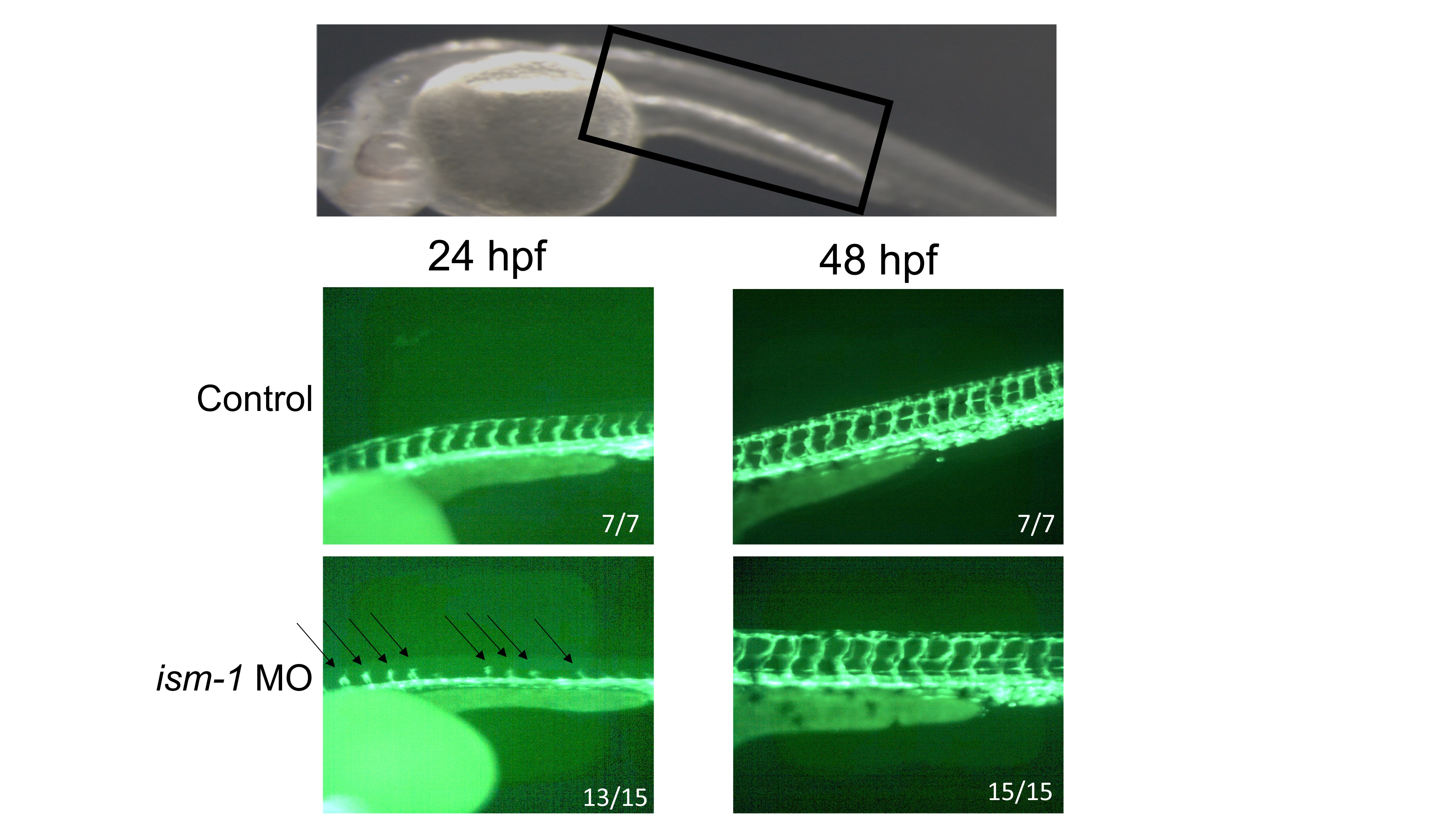
Figure Caption
Fig. S2
ism1 knockdown leads to temporally shortened intersegmental vessels.
flk1:GFP single-cell-stage embryos were injected with 7 ng of ism1 MO (bottom); uninjected embryos served as controls (top). 24 hpf (left column) and 48 hpf (right column) zebrafish were visualized at 40x for flk1:GFP fluorescence within the trunk area denoted by black box in brightfield image at top center. Arrows indicate shortened intersegmental vessels in ism1 morphants. Numbers in corner of images denote the number of embryos displaying the imaged phenotype.
Figure Data
Acknowledgments
This image is the copyrighted work of the attributed author or publisher, and
ZFIN has permission only to display this image to its users.
Additional permissions should be obtained from the applicable author or publisher of the image.
Full text @ PLoS One