
|
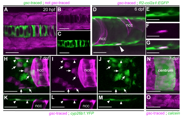
Fig. S1
Lineage tracing reveals that centra-associated col2a1- and cyp26b1-positive chordoblasts derive from the early notochord anlage. (A-C) Characterization of the gsc driver used for the axial mesoderm lineage tracing experiments. Dorsal view of a Tg(gsc:Cre); Tg(hsp70:loxp-dsRed-loxpegfp) double transgenic embryo at 15-somite stage (20 hours post fertilization; hpf), 2 hours post heatshock; confocal projection with merged red and green channels. (B) and (C) show the corresponding images of the single channels, respectively. Most notochordal cells express EGFP, whereas no EGFPpositive cells are present outside this structure, indicating the axial mesoderm specificity of the used gsc driver. In particular, sclerotomal cells of the somites are not labelled by the gsc driver. At the shown segmentation stage, notochordal cells have not segregated into outer chordoblasts and inner vacuolated cells as yet. Please note that compared to the endogenous gsc gene, the used gsc driver appears to lack negative cis-regulatory elements that usually repress expression within the notochord anlage, restricting expression to the prechordal plate (Doitsidou et al., 2002). (D) Lateral view of the notochord of a 6 dpf Tg(R2-col2a1a:EGFP); Tg(bact2:loxP-AmCyanloxP-mCherry) double transgenic larva injected with a gsc:Cre construct; confocal projection of merged green, red and transmitted light channels. Mosaic recombination events within the axial mesoderm result in mCherry labeling of some but not all cells deriving from the notochord anlage. The arrowhead points at an mCherry-expressing chordoblast identified as such by co-expression of the R2-col2a1a:EGFP transgene. (E-G) Magnification of the domain harboring the marked chordoblast from (D) in single and merged channels as indicated. In total, 28 chordoblasts positive for the gsc lineage tracer were observed in eight specimens. (H-M) Lateral view of the notochord of a 8 dpf Tg(cyp26b1:YFP); Tg(bact2:loxPAmCyan-loxPmCherry) double transgenic larva injected with the same gsc:cre construct; confocal projection of merged green and red (here magenta) channels (H,K) or the corresponding single channels (I,J,L,M); for better visualization, panels (K-M) show single confocal sections of the upper third from panels (H-J) with focus on marginal chordoblasts of the dorsal notochord border. Mosaic recombination within the axial mesoderm results in mCherry labeling of some chordoblasts as well as vacuolated notochordal cells (ncc). Some of the lineage-traced chordoblasts also coexpress the cyp26b1:YFP transgene (arrows and arrowheads), confirming their association with the centra inducing chordoblast subpopulation (seen in 11 centra of 4 specimens). mCherrypositive but YFP-negative chordoblasts most likely lie outside the RA target domain of the intercentral spaces. (N,O) Lateral view of a notochord at the level of a calcein-stained centrum (green) in a Tg(gsc:Cre); Tg(bact2:loxPAmCyan- loxP mCherry) double transgenic larva at 8dpf, confocal projection of merged red (here magenta) and green channels. Panel (O) shows the lower third of (N) in the single red (magenta) channel visualizing the continuous layer of notochord anlagen derived, mCherry-positive chordoblasts. Some of these cells are positioned directly underneath the mineralized matrix of the chordacentrum. Scale bars correspond to 20μm in (D-O) and to 50μm in (AC).