
|
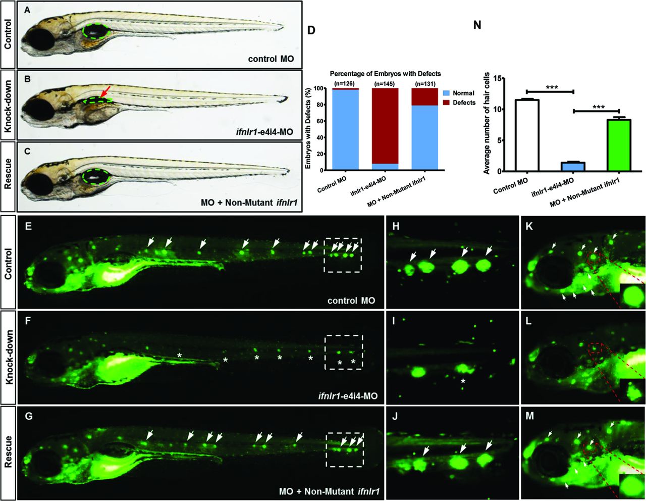
Fig. 4
Coinjection of ifnlr1 mRNA from non-mutant zebrafish rescued the phenotypes of non-inflated SB, hair cell loss and head neuromast loss (6 dpf) induced by ifnlr1-e4i4-MO. (A) Lateral views of control MO injected zebrafish embryos. (B) Embryos injected with ifnlr1 MO oligonucleotides (MO), ifnlr1 MO, plus non-mutant zebrafish ifnlr1. (C) Coinjection of non-mutant zebrafish ifnlr1 mRNA rescued non-inflated SB (red arrow) in ifnlr1 morphants at 6 dpf. (D) The bar graph shows the percentage of embryos with developmental defects. (E–N) Defective neuromasts in zebrafish. DASPEI (green) labelled neuromasts at 6 dpf. Neuromasts were stained as green dots (white arrow). The boxed regions are shown at higher magnification (right panels). (E,H,K) Control MO-injected zebrafish had normal numbers of hair cells. (F,I,L) Significantly decreased neuromast staining (asterisk) was observed in ifnlr1 morphants. (G,J,M) Coinjection of ifnlr1 mRNA from non-mutant zebrafish rescued ifnlr1-e4i4-MO from inducing hair cell loss and head neuromast loss (white arrow). Fluorescent DASPEI images were inverted for particle analysis. (N) The fluorescence particle signal in neuromasts was quantified using morphometric analysis. Quantification of the average number of neuromasts in the control embryos and the embryos injected with ifnlr1 MO or ifnlr1 MO plus non-mutant zebrafish ifnlr1. Error bars, SEM; ***P<0.0001 (n=10; ANOVA). DASPEI, 2-(4-(dimethylamino)styryl)-N-ethylpyridinium iodide; MO, Morpholino; SB, swim bladder.