
IMAGE
Fig. 3
- ID
- ZDB-IMAGE-180717-3
- Publication
- Yoo et al., 2018 - Impact of Nicotine Exposure on Hair Cell Toxicity and Embryotoxicity During Zebrafish Development
- All Figures
- Figures for Yoo et al., 2018
Image
|
Figure Caption
Fig. 3
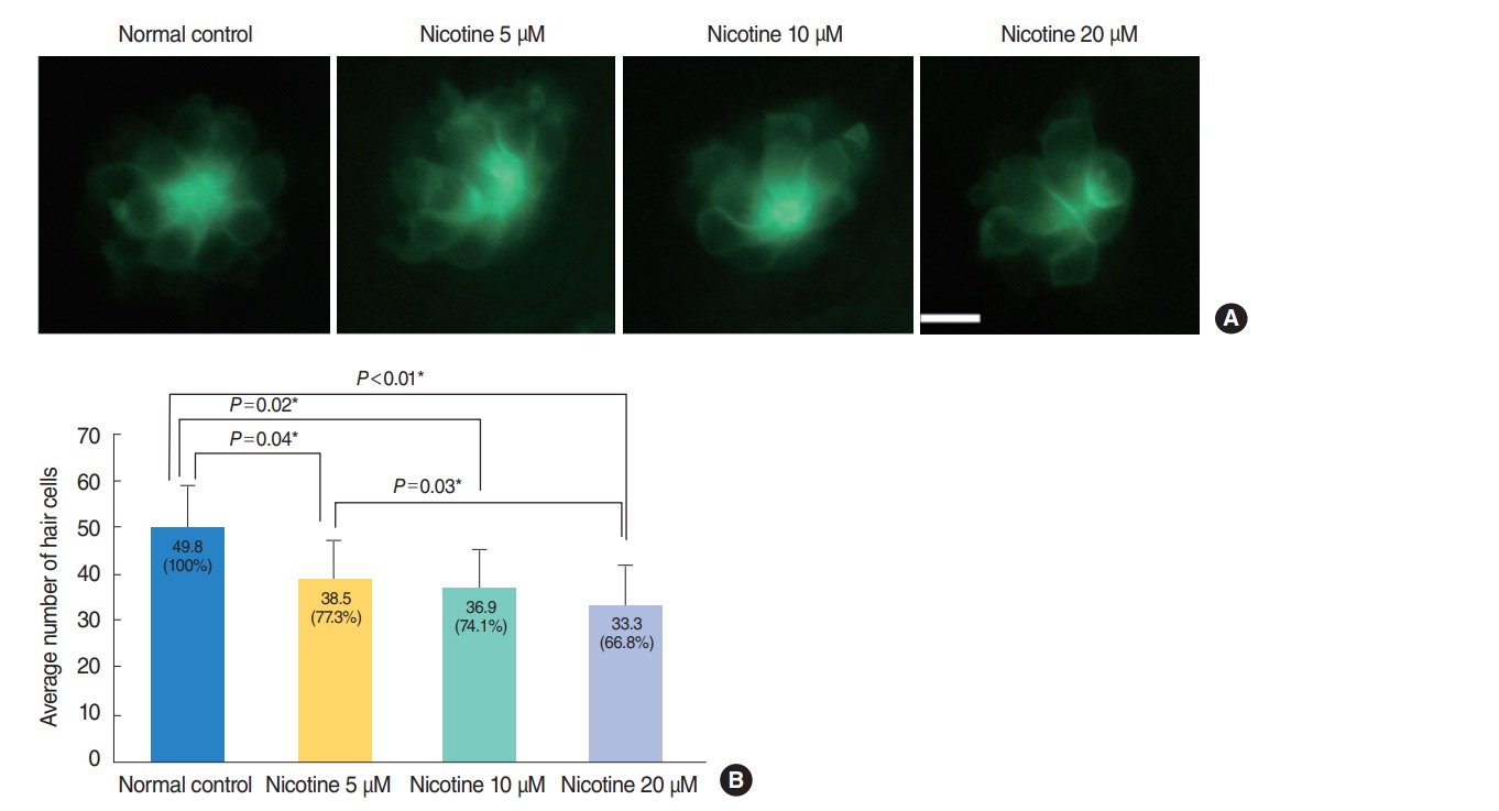
Nicotine-induced hair cell toxicity at 120 hours post-fertilization (hpf). The total number of hair cells was measured in four neuromasts (supraorbital [SO1 and SO2], otic [O1], and occipital [OC1]; total n=150). (A) The number of hair cells decreased in nicotine-exposed groups as shown by fluorescent microscopy (OC1, ×40). Scale bar=10 μm (×10). (B) Animals in the normal control group had an average of 49.8 hair cells. Nicotine exposure significantly decreased the number of total hair cells of the four neuromasts compared with that of the normal control, (P<0.001, one-way analysis of variance). At a concentration of 40 μM nicotine almost all the fish were dead, therefore statistical analysis was unavailable. All data were evaluated using transgenic (brn3c:EGFP) zebrafish at 120 hpf. Only statistically significant pair-wise comparisons in posthoc analysis are shown. *Statistically significant (P<0.05).
Acknowledgments
This image is the copyrighted work of the attributed author or publisher, and
ZFIN has permission only to display this image to its users.
Additional permissions should be obtained from the applicable author or publisher of the image.
Full text @ Clin. Exp. Otorhinolaryngol.