
Image
|
Figure Caption
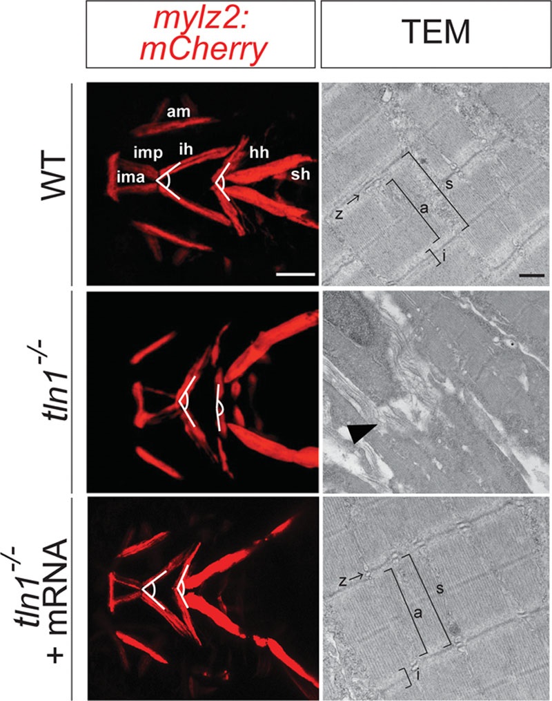
Fig. 5
Defects in craniofacial skeletal muscles in tln1 mutants. Confocal images of craniofacial skeletal muscles in Tg(mylz2:mCherry) transgenic reveal increased intermuscular tension in the craniofacial muscles in the mutants (left and center) when compared with WT (left and above). TEM images of the transverse section of the ih (depicted by white dotted line, above and center) show complete disruption of the sarcomere structure in the mutants, depicted by black arrowhead (right and center) when compared with WT (right and above). The Z-line and the A-and I-bands, observed in the WT (right and above) are completely absent in the mutants (right and center). Injection of full-length tln1 mRNA partially rescues the phenotypes as observed by decreased intermuscular tension in the craniofacial muscles in the rescued mutants (left and below). TEM images of the transverse section of the ih in the rescued mutants show reorganized sarcomere structure, similar to the WT (right and below). Scale bar: 500μm (Tg(mylz2: mCherry)) and 500nm (TEM). a, A-band; am, adductor mandibulae; i, I-band; ima, intermandibularis anterior; imp, intermandibularis posterior; s, sarcomere unit; sh, sternohyoideus.
Figure Data
Acknowledgments
This image is the copyrighted work of the attributed author or publisher, and
ZFIN has permission only to display this image to its users.
Additional permissions should be obtained from the applicable author or publisher of the image.
Full text @ Plast Reconstr Surg Glob Open