
|
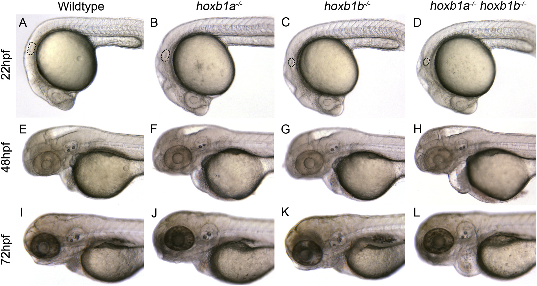
Fig. S1
Morphology of paralog group 1 hox mutants.
Embryos were imaged consecutively at 22hpf (A–D), 48hpf (E-H) and 72hpf (I–L) to examine gross morphological defects. Hoxb1a−/− mutants (B, F, J) do not have any major morphological defects as compared to wildtype embryos. (A, E, I) Hoxb1b−/− mutants have a small otic vesicle at 22hpf (C) which does not persist at later stages (G, K). Hoxb1a−/− hoxb1b−/− mutants have a small otic vesicle at 22hpf (D) and develop cardiac edema at later stages (H, L). All embryos have been genotyped for hoxb1asa1191 and hoxb1bua1006. All embryos are lateral views, anterior to the left. Hpf, hours post fertilization.
Reprinted from Mechanisms of Development, 150, Selland, L.G., Koch, S., Laraque, M., Waskiewicz, A.J., Coordinate regulation of retinoic acid synthesis by pbx genes and fibroblast growth factor signaling by hoxb1b is required for hindbrain patterning and development, 28-41, Copyright (2018) with permission from Elsevier. Full text @ Mech. Dev.