
|
Fig. 4
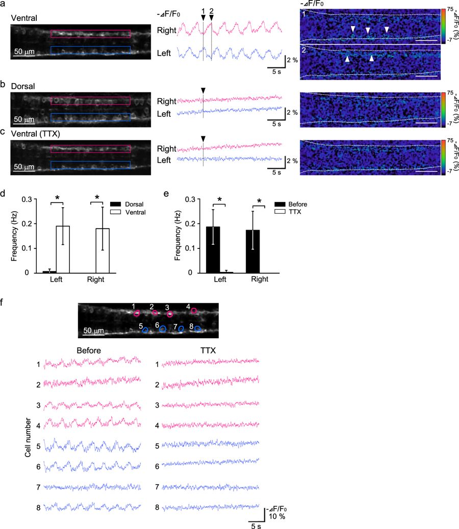
Spontaneous activity of spinal cord neurons was detected via ASAP1 imaging. (a) Changes in ASAP1 fluorescence were observed in the ventral region of the spinal cord. (left) Dorsal view of the ventral spinal cord of Tg (elavl3:GAL4-VP16;UAS:ASAP1) fish. The rostral side is to the left, and the area between 3–8 somites is shown. Regions of interest (ROIs) located between 5–7 somites are indicated by red (right side) and blue (left side) rectangles. (middle, right) Fluorescence changes of ASAP1 in the ROIs are shown in the middle panel. The images at two time points indicated by black arrowheads are shown in the right panel. White arrowheads indicate the activated neurons. Changes in fluorescence (−ΔF/F0) are indicated by the pseudocolor scale shown at right. (b) Fluorescence changes of ASAP1 were not observed in the dorsal region of the spinal cord. (left) Dorsal view of the same embryo shown in (a) with the image focus at the ventral region. An image at the time point indicated by the black arrowhead in the middle panel is shown to the right. (c) The spontaneous activity in the ventral region of the spinal cord was reduced by tetrodotoxin (TTX) treatment. The imaged plane and ROIs are in the same positions as in (a). (d) The frequency of the fluorescence changes was significantly higher in the ventral region. Similar results were obtained for both the left and right sides of the spinal cord (6 fish, *p < 0.05). (e) The spontaneous activity was almost eliminated by TTX treatment (6 fish, *p < 0.05). (f) Monitoring of spontaneous activity of individual cells (cells 1–8) by ASAP1 imaging. (top) A fluorescence image of the ventral spinal cord of Tg(elavl3:GAL4-VP16;UAS:ASAP1) fish. ROIs were located at the 8 cells (red: right side, blue: left side). Examples of activity patterns of the 8 cells for two conditions (before and after TTX treatment) are shown in the lower panel.