
|
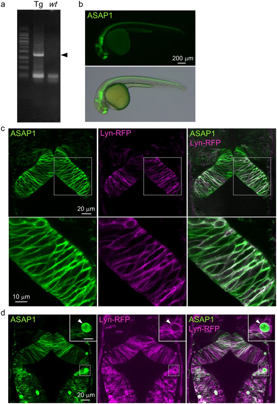
Fig. 2
Transgenic zebrafish showed membrane-localized ASAP1 in the neural tube. (a) Genotyping results of Tg(UAS:ASAP1) fish. Left: Tg(UAS:ASAP1) (Tg), right: wild-type (wt). The cropped gel image is shown. The full-length gel is presented in Supplementary Fig. S6a. (b) ASAP1 was distributed widely in the neural tube of Tg(elavl3:GAL4-VP16;UAS:ASAP1) fish at 1 dpf. (c) Dorsal view of the neural tube of Tg(elavl3:GAL4-VP16;UAS:ASAP1;UAS:lyn-RFP) embryos at 1 dpf. ASAP1 (green) was co-localized with Lyn-RFP (red: cell membranes) in the neural tube. Higher magnification images are shown in the lower panel. (d) Some of the ASAP1 positive cells had irregular shapes (arrowheads). Higher magnification images are shown in the upper right corner.