
|
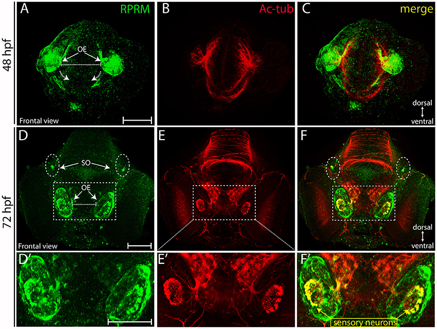
Fig. 4
Expression of RPRM in zebrafish olfactory epithelium. RPRM protein expression was analyzed by IF in wild-type embryos. (A–F) Frontal views with dorsal to the top and ventral to the bottom (double arrows in A,D). At 48 hpf (A–C) RPRM is expressed in the OE showing co-localization with Ac-tub. (B–C). At 72 hpf, (D–F) RPRM is expressed at the OE and co-localizes with olfactory sensory neurons (OSNs) which express Ac-tub (F–F′) and some of the axons projecting to the OB, also is present in the supraorbital neuromasts (SO) located bilaterally (dotted circles in D, F). (D′–F′) inset magnification at the OE showing RPRM and Ac-tub expression in OSNs (yellow square and arrows). Scale Bars in (A,D,D′): 100 μm.