
|
Fig. S1
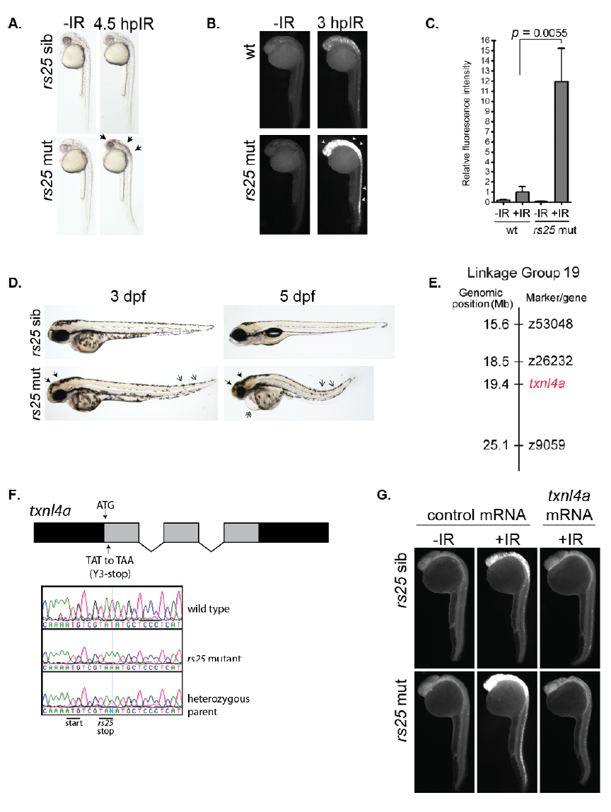
The rs25 mutation disrupts the txnl4a gene (A) rs25 siblings and mutants were exposed to 10 Gy IR and analyzed by bright field microscopy at 4.5 hpIR. Black arrows in the rs25 mutants mark the accumulation of cell death. (B-C) Wild type embryos and rs25 mutants were exposed to 10 Gy IR at 24 hpf, fixed at 3 hpIR, analyzed by active Caspase-3 and fluorescence was quantified. White arrowheads in the rs25 mutants mark the accumulation of active Caspase-3-marked apoptosis. (D) rs25 siblings and mutants were imaged by bright field microscopy at 3 and 5 dpf. Solid arrowheads in rs25 mutants mark accumulation of cell death in the head. Single, open arrowheads mark the curved tail, and double arrowheads mark heart edema. (E) The rs25 mutation was localized to linkage group 19 between flanking markers z26232 and z9059. (F) Sequencing of rs25 mutants revealed a thymine to adenine transition within the txnl4a coding sequence replacing the third amino acid (tyrosine) with a stop codon (Y3-Stop). Schematic of txnl4a shows 3 exons (in grey boxes), the 5’ and 3’ untranslated region (in black boxes), and introns (bent lines). Sequencing of the txnl4a gene is shown for wild type, rs25 mutant, and an rs25 heterozygote. A vertical blue line intersects the position of the mutated nucleotide. The solid, black lines under the sequencing designate the start of the coding sequence and rs25-mediated premature stop codon. (G) Embryos from an incross of rs25 heterozygotes were injected with control mRNA (GFP) or txnl4a mRNA. Embryos were exposed (or left unexposed) to 10 Gy at 24 hpf and analyzed 3 hours later for active Caspase-3.