
Image
|
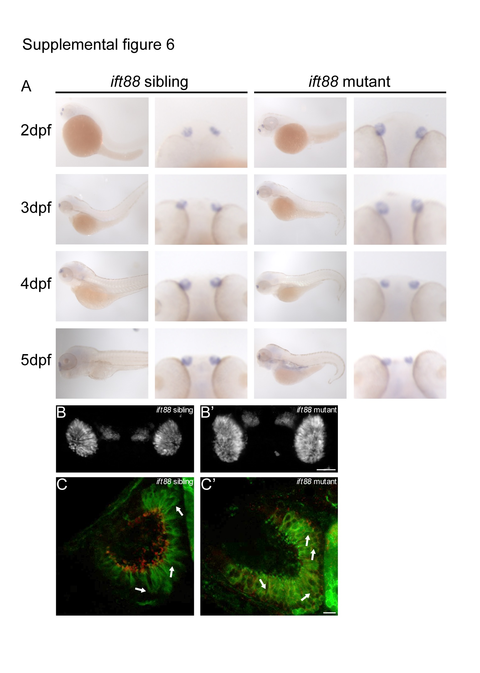
Figure Caption
Fig. S6
Ciliated OSNs stained with anti-GFP (green) and anti-G /olf (red) present in both sibling and ift88 mutant. Lower panels: the anti-G /olf (red) signal only, demonstrating presence of anti-G /olf (red) staining in the cell bodies of both sibling and ift88 mutant. Scale bar is 10 µm.
Acknowledgments
This image is the copyrighted work of the attributed author or publisher, and
ZFIN has permission only to display this image to its users.
Additional permissions should be obtained from the applicable author or publisher of the image.
Full text @ Cilia