
Image
|
Figure Caption
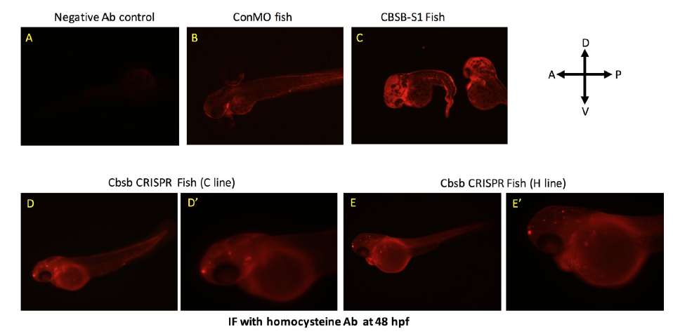
Fig. S7
Homocysteine IF in cbsb morphant and CRISPR fish. IF with homocyteine antibody was performed in 48 hpf control MO (ConMO) fish (A) and cbsb splice1 MO (CBSB-S1) injected (C) fish. Panel A shows fish with no primary antibody control. D and E are cbsb CRISPR 48 hpf fish from C and H line respectively. D’ and E’ are high power images of the head and yolk region. Note the halo staining around the yolk, which is indicative of homocysteine proteins in yolk as previously reported (Matthews et al., 2009).
Figure Data
Acknowledgments
This image is the copyrighted work of the attributed author or publisher, and
ZFIN has permission only to display this image to its users.
Additional permissions should be obtained from the applicable author or publisher of the image.
Full text @ Front Cell Dev Biol