
|
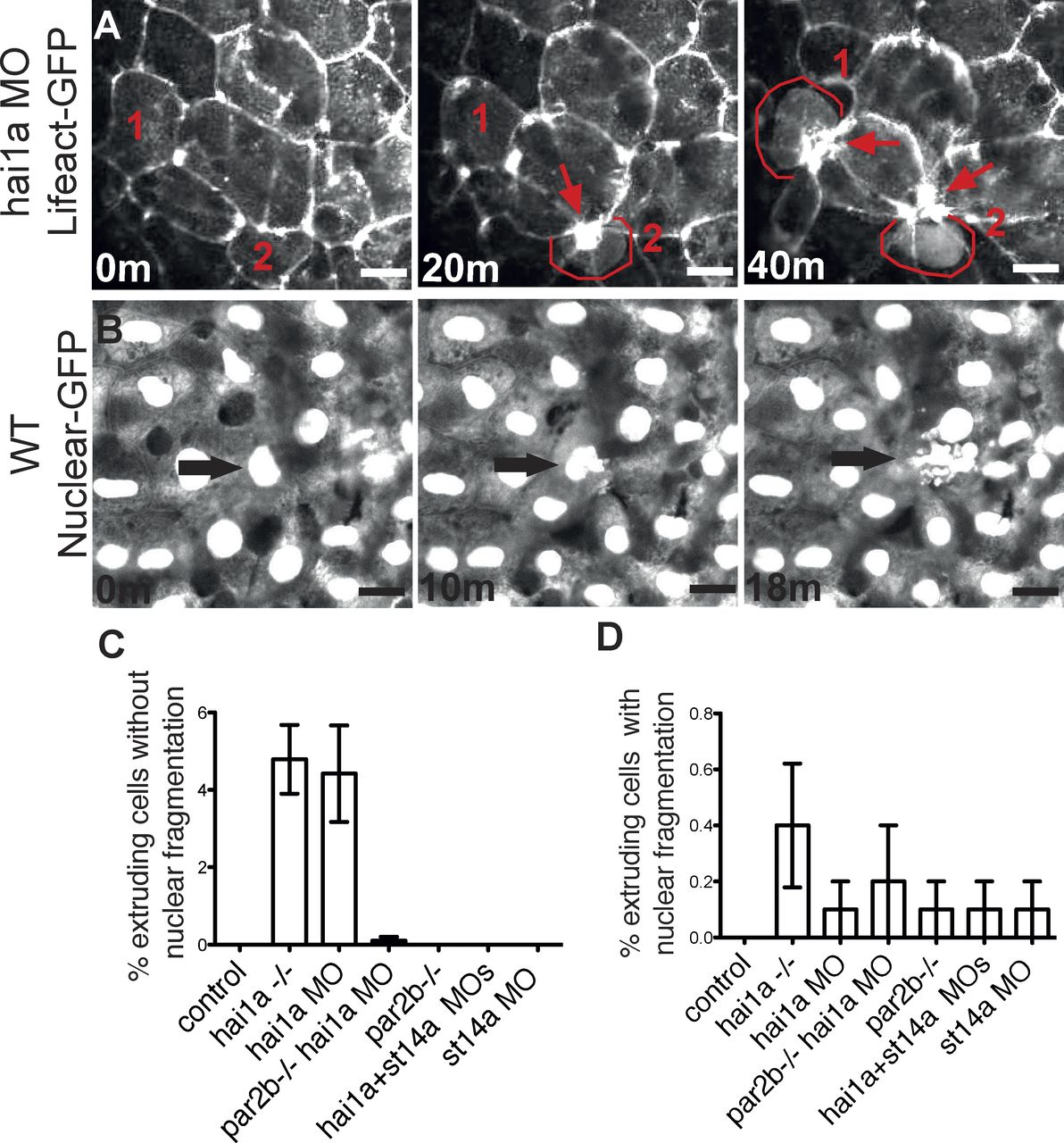
Fig. 3
par2b-dependent apical extrusion of epithelial cells from periderm in hai1a morphant embryos. (A) The yolk sac periderm of a hai1a morphant periderm-LifeActGFP embryo was imaged from 28 to 46 hpf. Three time-lapse panels spanning 40 min show an F-actin ring that contracts around cells being extruded apically from the periderm. Cells destined to extrude are labeled 1 and 2 in all panels and are highlighted with red outline after extrusion has begun. Contracted actin rings remaining at the site of extrusion and at the center of rosettes formed by surrounding cells are marked by red arrows. Bar, 5 µm. (B) The yolk sac periderm of a control periderm-nuclear-GFP embryo was imaged from 28 to 46 hpf. Three panels spanning 18 min show extrusion of a cell with an obviously fragmented nucleus (arrow). Bar, 5 µm. (C and D) Quantitation of extrusion events in control and hai1a morphants with or without Par2b or matriptase (st14a) deficiency. The ventral yolk sac of 10 periderm-nuclear-GFP embryos for each condition was imaged from 28 to 46 hpf, and extrusion events were counted and classified by whether the nucleus of the extruding cell was not (C) or was (D) obviously fragmented. Results are expressed as the number of extrusion events as a percentage of the number of cells, usually ∼200, present in the field at the start of the imaging period (mean ± SEM; n = 10). The hai1a morphant and mutant groups in C were different from all other groups by one-way ANOVA and Bonferroni posttest, P < 0.0001. No groups in D were different from control.