
|
Fig. S2
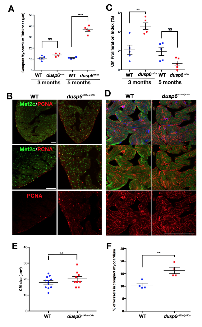
dusp6 mutant heart are larger and have a thicker compact myocardium containing more vessels than WT hearts (A) Quantification of compact myocardium thickness in uninjured hearts at 3 and 5 months of age in WT and dusp6 mutant (n≥4 for each group). At 5 months of age dusp6 mutant fish have thicker compact myocardium than WT hearts. ****p<0.0001; ns= not significant. One-way ANOVA. (B) 3 months old uninjured zebrafish hearts immunostained for Mef2c (green; cardiomyocyte nuclei) and PCNA (red; proliferation marker). dusp6 heart (n=5) have more proliferating cardiomyocyte than WT hearts (n=5). (C) Quantification of cardiomyocyte proliferation in WT and dusp6 mutant hearts at 3 and 5 months of age (n=5 for each group). **p<0.01; ns= not significant. One-way ANOVA. (D) Uninjured hearts sections stained for MHC to visualize cardiomyocyte and wheat germ agglutinin (WGA) to stain cell membrane. (E) Quantification of cardiomyocyte size in uninjured hearts. dusp6 mutant hearts (n=9) have the same size of cardiomyocyte compared to WT (n=11); Student’s t-test. n.s.=not significant. (F) Quantification of vessels area per compact myocardium area in uninjured hearts at 5 months of age WT (n=4) and dusp6 mutant (n=4). **p<0.01. Student’s t-test. Scale bars, 100 μm.