
Image
|
Figure Caption
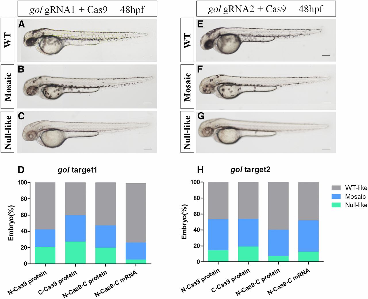
Fig. 6
Mutagenetic phenotype of various nuclear localization signal (NLS)-fused Cas9 proteins and N-Cas9-C mRNA for two sites of the gol gene. (A–C) and (E–G) show lateral views of wild-type (WT) (A and E), gol-target 1 (B and C), and gol-target 2 (F and G) at 48 hpf (hr postfertilization). The phenotypes observed were WT (A and E), mosaic retinal pigmented epithelium (RPE) (B and F), and unpigmented RPE (C and G). (D and H) Proportions of embryos with each phenotype. Scale bars: 0.2 mm.
Acknowledgments
This image is the copyrighted work of the attributed author or publisher, and
ZFIN has permission only to display this image to its users.
Additional permissions should be obtained from the applicable author or publisher of the image.
Full text @ G3 (Bethesda)