
|
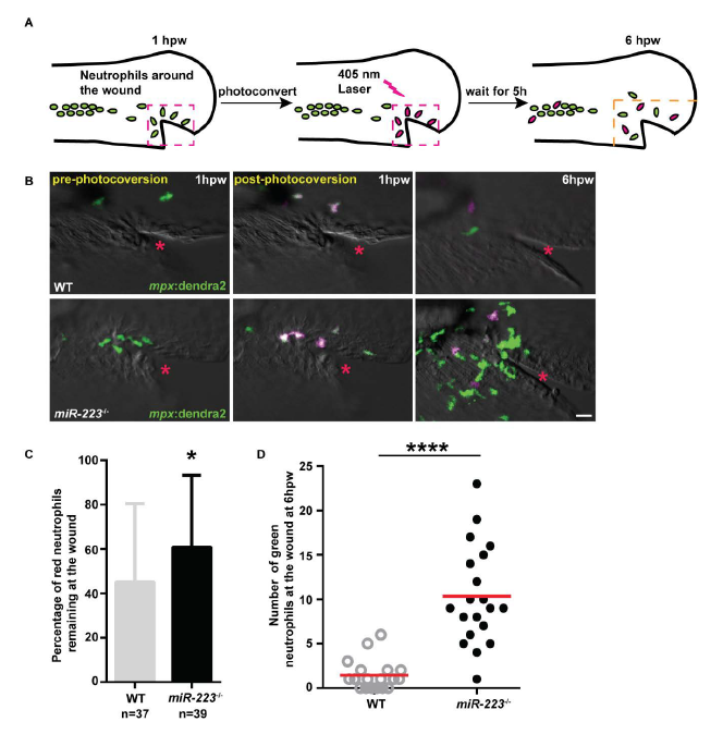
Fig. S2
Enhanced neutrophil recruitment in miR-223-/- embryos results from both continuous neutrophil recruitment and a defect in neutrophil reverse migration, related to Figure 1. (A) Schematics of photoconversion-enabled neutrophil fate-mapping assay. Embryos from Tg(miR-223-/-, mpx: Dendra2) or Tg(mpx: Dendra2) were used. Photolabled neutrophils remaining at the wound indicate a defect in reverse migration. (B) Representative confocal images of embryos at three time points: before, immediately after, and 5 hours after photoconversion. Red neutrophils are photoconverted neutrophils. Red star indicates the injury site. (C) Percentage of red neutrophils remaining at the wound at 6 hpw. (D) Quantification of the number of green neutrophils at the wound at 6 hpw. Scale bars, 20 μm. * P < 0.05 and **** P < 0.0001, unpaired student t-test.