
|
Fig. 2
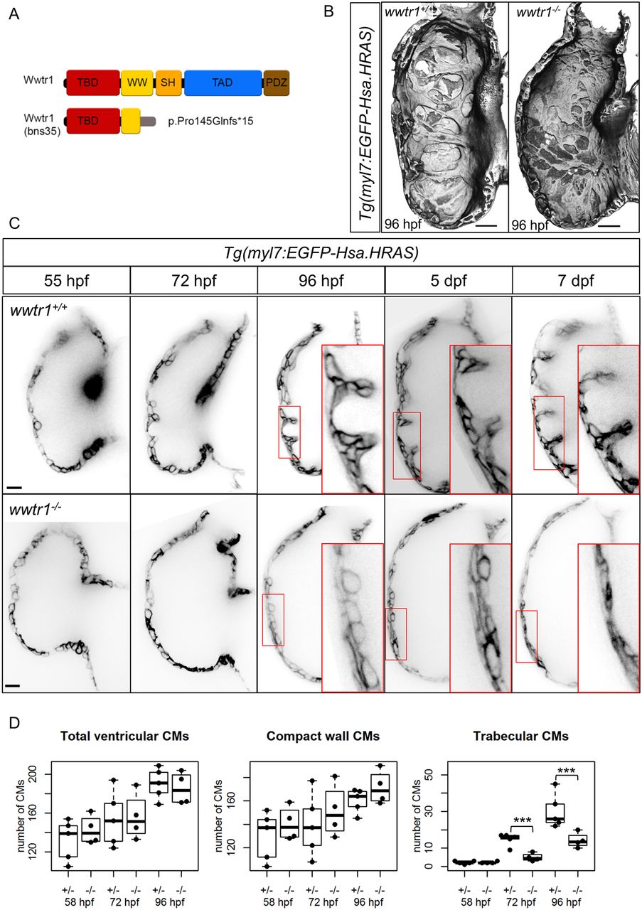
wwtr1 mutant hearts do not develop trabecular ridges. (A) Schematic of wild-type and predicted truncation of Wwtr1 due to a CRISPR-induced out-of-frame insertion (see Materials and Methods). (B) 3D surface reconstruction of ventricular chambers at 96 hpf shows distinct and prominent muscular ridges in wwtr1+/+ (8/8) and wwtr1+/− (7/7), but not wwtr1−/− (20/24), hearts. Scale bars: 15 μm. (C) Confocal sagittal sections of wild-type and wwtr1−/− ventricular chambers of the same animal from 55 hpf to 7 dpf. Scale bars: 15 μm. (D) Longitudinal quantification of the number of ventricular cardiomyocytes (CMs) from five wwtr1+/− and four wwtr1−/− hearts. The total number of ventricular CMs is divided into two groups: compact wall CMs and trabecular CMs. Each point represents a heart. ***P<0.001 by Poisson regression. The box covers the inter-quartile range (IQR); the whiskers cover the first and last quartile of the data. Data points outside the whiskers are outliers (>1.5×IQR from the first and third quartile).