
|
Fig. S2
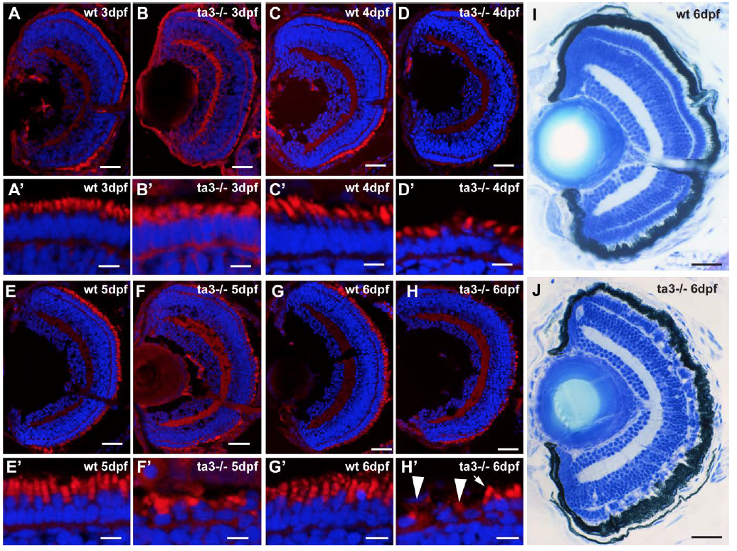
Time course of PR degeneration in ta3 mutants
Retinal cryosections of wt and ta3-/- zebrafish at 3dpf (A-B'), 4dpf (C-D'), 5dpf (E-F') and 6 dpf (G-H'). Nuclei are counterstained with DAPI and membranes (including outer segments) are highlighted with BODIPY. Note the progressive cell shape loss and decreased numbers of outer segments in mutants. The arrow in H' indicates a PR with an OS, while the arrowheads point to PRs without OSs. (I-J) Histological sections through whole wt and ta3-/- eyes at 6dpf showing gaps in the PR cell layer, but also persistance of many PR cells. Scale bars: 30 μm in (A-H) and (I-J), 4 μm in A'-H'.